Skip to Content
Merck
All Photos(1)

Key Documents

346M-1

Sigma-Aldrich

Villin (CWWB1) Mouse Monoclonal Antibody

Sign Into View Organizational & Contract Pricing


About This Item

UNSPSC Code:
12352203
NACRES:
NA.41
clone:
CWWB1, monoclonal
application:
IHC (p)
species reactivity:
human
technique(s):
immunohistochemistry (formalin-fixed, paraffin-embedded sections): 1:100-1:500
citations:
7

biological source

mouse

Quality Level

100
500

conjugate

unconjugated

antibody form

diluted ascites fluid

antibody product type

primary antibodies

clone

CWWB1, monoclonal

description

For In Vitro Diagnostic Use in Select Regions (See Chart)

form

buffered aqueous solution

species reactivity

human

packaging

vial of 0.1 mL concentrate (346M-14)
vial of 0.5 mL concentrate (346M-15)
bottle of 1.0 mL predilute (346M-17)
vial of 1.0 mL concentrate (346M-16)
bottle of 7.0 mL predilute (346M-18)

manufacturer/tradename

Cell Marque

technique(s)

immunohistochemistry (formalin-fixed, paraffin-embedded sections): 1:100-1:500

isotype

IgG1

control

colon

shipped in

wet ice

storage temp.

2-8°C

visualization

cytoplasmic

Gene Information

human ... VIL1(7429)

General description

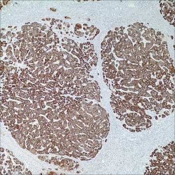
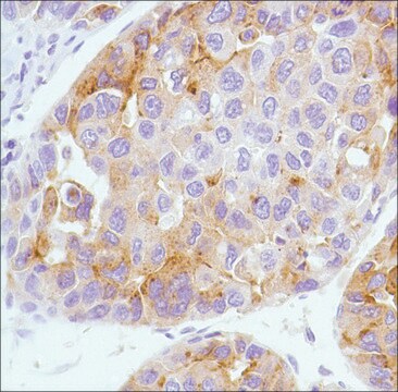
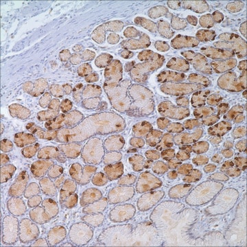
Villin is a 95 kD glycoprotein of microvilli associated with rootlet formation in gastrointestinal mucosal epithelium. Anti-villin labels the brush border area in the gastrointestinal mucosal epithelium. This antibody has been useful in differentiating gastrointestinal adenocarcinoma, neuroendocrine carcinomas, and ovarian adenocarcinomas from adenocarcinomas of other organs. This antibody also labels Merkel cells of the skin.

Quality


IVD

IVD

IVD

RUO

Linkage

Villin Positive Control Slides, Product No. 346S, are available for immunohistochemistry (formalin-fixed, paraffin-embedded sections).

Physical form

Solution in Tris Buffer, pH 7.3-7.7, with 1% BSA and <0.1% Sodium Azide

Preparation Note

Download the IFU specific to your product lot and formatNote: This requires a keycode which can be found on your packaging or product label.

Other Notes

For Technical Service please contact: 800-665-7284 or email: service@cellmarque.com

Legal Information

Cell Marque is a trademark of Merck KGaA, Darmstadt, Germany

Not finding the right product?  

Try our Product Selector Tool.


Choose from one of the most recent versions:

Certificates of Analysis (COA)

Lot/Batch Number

Don't see the Right Version?

If you require a particular version, you can look up a specific certificate by the Lot or Batch number.

Already Own This Product?

Find documentation for the products that you have recently purchased in the Document Library.

Visit the Document Library

Sean K Lau et al.
Human pathology, 33(12), 1175-1181 (2003-01-07)
Distinguishing hepatocellular carcinoma (HCC) from cholangiocarcinoma (CC) and metastatic adenocarcinoma (MA) involving the liver can be problematic, often requiring the use of immunohistochemistry to facilitate diagnosis. Hep Par 1, a monoclonal antibody with expression confined primarily to benign and malignant
P J Zhang et al.
Archives of pathology & laboratory medicine, 123(9), 812-816 (1999-08-24)
Villin, a 95-kd cytoskeletal protein associated with axial microfilament bundles of brush border microvilli, is mostly restricted to intestinal glandular tumors. Villin immunoexpression was recently observed in a small number of carcinoids of the intestinal tract and lung, but its
Satoshi Nishizuka et al.
Cancer research, 63(17), 5243-5250 (2003-09-23)
Colon and ovarian cancers can be difficult to distinguish in the abdomen, and the distinction is important because it determines which drugs will be used for therapy. To identify molecular markers for that differential diagnosis, we developed a multistep protocol
N S Goldstein et al.
American journal of clinical pathology, 116(3), 319-325 (2001-09-14)
We studied 14 mucinous and 26 nonmucinous bronchioloalveolar adenocarcinomas (BACs) with thyroid transcription factor (TTF), cytokeratin (CK) 7, CK20, and villin to characterize their staining patterns with these antibodies and identify staining differences between the neoplasms. We also stained 11
Pheroze Tamboli et al.
Archives of pathology & laboratory medicine, 126(9), 1057-1063 (2002-09-03)
To determine whether a limited immunohistochemical panel can help differentiate metastatic colonic adenocarcinoma from primary enteric-type adenocarcinoma of the urinary tract and urothelial (transitional cell) carcinoma with glandular differentiation, which appear morphologically similar but most often necessitate different treatment protocols.

Articles

Colorectal cancer is the third most common cancer in both men and women. An estimated 136,000 cases of colorectal cancer are expected to occur in 2016.

Our team of scientists has experience in all areas of research including Life Science, Material Science, Chemical Synthesis, Chromatography, Analytical and many others.

Contact Technical Service