Skip to Content
Merck
All Photos(2)

Key Documents

C2687

Sigma-Aldrich

Monoclonal Anti-Calponin antibody produced in mouse

clone hCP, ascites fluid

Synonym(s):

Anti-Calponin-1

Sign Into View Organizational & Contract Pricing


About This Item

MDL number:
UNSPSC Code:
12352203
NACRES:
NA.41

biological source

mouse

Quality Level

conjugate

unconjugated

antibody form

ascites fluid

antibody product type

primary antibodies

clone

hCP, monoclonal

mol wt

antigen 34 kDa

contains

15 mM sodium azide

species reactivity

rat, rabbit, pig, human, mouse

technique(s)

immunohistochemistry (formalin-fixed, paraffin-embedded sections): suitable
immunoprecipitation (IP): suitable
microarray: suitable
western blot: 1:10,000 using human uterus extract

isotype

IgG1

UniProt accession no.

shipped in

dry ice

storage temp.

−20°C

target post-translational modification

unmodified

Gene Information

Related Categories

Specificity

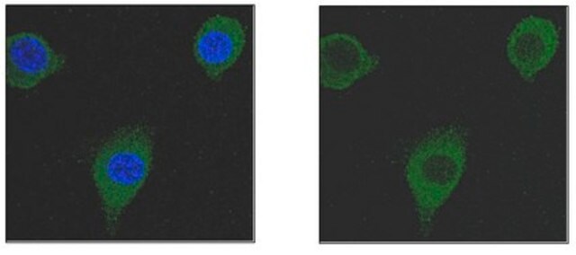
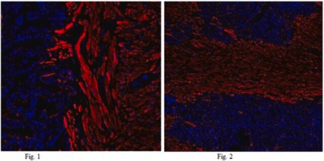
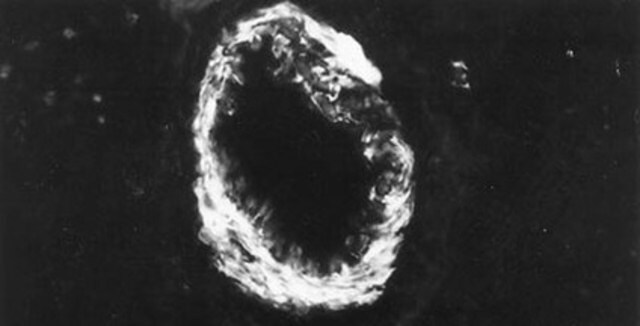
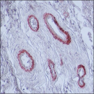
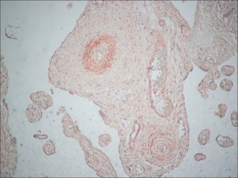
The antibody hCP (also cited as CALP), localizes calponin in mammalian smooth muscle. In human uterus, an additional band of approximately 27 kDa (l-calponin) may also be stained. The antibody does not cross-react with skeletal, cardiac or non-muscle tissue calponin. Nevertheless, the antibody exhibits cross-reactivity with an epitope (150 kDa range) in human or mouse skeletal muscle. In immunohistochemical staining, the product exhibits smooth muscle specificity. It stains vascular and visceral smooth muscle cells in tissue sections and primary cultured cells (or early passages), but not most cell lines originally derived from smooth muscle. The antibody does not stain epithelial, endothelial or connective tissue fibroblast cells. This product does not cross react with smooth muscle tissue from chicken.

Immunogen

human uterus smooth muscle extract.

Application

Monoclonal Anti-Calponin antibody produced in mouse is suitable for the following applications:
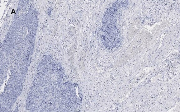
  • Immunohistochemistry using formalin-fixed, paraffin-embedded sections
  • Immunoprecipitation
  • Microarray
  • Western blotting (at a dilution of 1:10,000 using human uterus extract)
  • Flow cytometry analysis

Biochem/physiol Actions

Calponin is a calcium binding protein that belongs to a family of actin-associated proteins. Calponin is necessary for autophosphorylation of protein kinase C (PKC) in vascular smooth muscle (VSM). Calponin-1 inhibition can prevent uterine smooth muscle cell migration, cause morphological change and rearrange F-actin without affecting its proliferation and apoptosis. Calponin h1 (CN) is a differentiation marker of smooth muscle cells and is down-regulated in the blood vessels of several human tumors. It can inhibit actomyosin ATPase activity. The h1 and h2 calponins bind F-actin and play a key role in regulating actin filaments in smooth muscle and non-muscle cells.

Disclaimer

Unless otherwise stated in our catalog or other company documentation accompanying the product(s), our products are intended for research use only and are not to be used for any other purpose, which includes but is not limited to, unauthorized commercial uses, in vitro diagnostic uses, ex vivo or in vivo therapeutic uses or any type of consumption or application to humans or animals.

Not finding the right product?  

Try our Product Selector Tool.

Storage Class Code

10 - Combustible liquids

WGK

WGK 1

Flash Point(F)

Not applicable

Flash Point(C)

Not applicable


Certificates of Analysis (COA)

Search for Certificates of Analysis (COA) by entering the products Lot/Batch Number. Lot and Batch Numbers can be found on a product’s label following the words ‘Lot’ or ‘Batch’.

Already Own This Product?

Find documentation for the products that you have recently purchased in the Document Library.

Visit the Document Library

A Draeger et al.
FEBS letters, 291(1), 24-28 (1991-10-07)
Two-dimensional gel analysis of basic proteins in developing human smooth muscle identifies calponin as a prominent marker of the differentiated phenotype. Adult tissue (human and mouse) typically expresses up to four calponin isoforms, three of which appear sequentially during fetal
M G Frid et al.
Developmental biology, 153(2), 185-193 (1992-10-01)
Expression of the regulatory contractile proteins, heavy caldesmon (h-caldesmon) and calponin was studied in human aortic smooth muscle cells (SMCs) during development and compared with the expression of alpha-SM-actin and smooth muscle-myosin heavy chain (SM-MHCs). For this study, novel monoclonal
Won Sun Park et al.
American journal of physiology. Cell physiology, 305(4), C377-C391 (2013-06-14)
Human adipose tissue-derived mesenchymal stem cells (hASCs) have the power to differentiate into various cell types including chondrocytes, osteocytes, adipocytes, neurons, cardiomyocytes, and smooth muscle cells. We characterized the functional expression of ion channels after transforming growth factor-β1 (TGF-β1)-induced differentiation
Gaëlle Pérot et al.
Cancer research, 69(6), 2269-2278 (2009-03-12)
Myocardin (MYOCD), a serum response factor (SRF) transcriptional cofactor, is essential for cardiac and smooth muscle development and differentiation. We show here by array-based comparative genomic hybridization, fluorescence in situ hybridization, and expression analysis approaches that MYOCD gene is highly
Mun Chun Chan et al.
Molecular and cellular biology, 31(3), 517-530 (2010-12-08)
Pulmonary artery hypertension (PAH) is characterized by elevated pulmonary artery resistance and increased medial thickness due to deregulation of vascular remodeling. Inactivating mutations of the BMPRII gene, which encodes a receptor for bone morphogenetic proteins (BMPs), are identified in ∼60%

Our team of scientists has experience in all areas of research including Life Science, Material Science, Chemical Synthesis, Chromatography, Analytical and many others.

Contact Technical Service