Skip to Content
Merck
All Photos(17)

Key Documents

ZRB1838

Sigma-Aldrich

Anti-Phospho-EGFR (Tyr1068) Antibody, clone 3F4 ZooMAb® Rabbit Monoclonal

enhanced validation
greener alternative

recombinant, expressed in HEK 293 cells

Synonym(s):

EC:2.7.10.1, Epidermal growth factor receptor, Proto-oncogene c-ErbB-1, Receptor tyrosine-protein kinase erbB-1

Sign Into View Organizational & Contract Pricing


About This Item

UNSPSC Code:
12352203
NACRES:
NA.41
conjugate:
unconjugated
application:
FACS
ICC
IHC
WB
affinity binding assay
clone:
3F4, recombinant monoclonal
species reactivity:
human
citations:
technique(s):
affinity binding assay: suitable
flow cytometry: suitable
immunocytochemistry: suitable
immunohistochemistry: suitable
western blot: suitable

biological source

rabbit

Quality Level

recombinant

expressed in HEK 293 cells

conjugate

unconjugated

antibody form

purified antibody

antibody product type

primary antibodies

clone

3F4, recombinant monoclonal

description

3F4 Clone

product line

ZooMAb® learn more

form

lyophilized

mol wt

calculated mol wt 134.28 kDa
observed mol wt ~160 kDa

purified by

using Protein A

species reactivity

human

species reactivity (predicted by homology)

monkey

packaging

antibody small pack of 25 μL

greener alternative product characteristics

Waste Prevention
Designing Safer Chemicals
Design for Energy Efficiency
Learn more about the Principles of Green Chemistry.

enhanced validation

recombinant expression
Learn more about Antibody Enhanced Validation

technique(s)

affinity binding assay: suitable
flow cytometry: suitable
immunocytochemistry: suitable
immunohistochemistry: suitable
western blot: suitable

isotype

IgG

epitope sequence

C-terminal

Protein ID accession no.

UniProt accession no.

greener alternative category

shipped in

ambient

storage temp.

2-8°C

Gene Information

human ... EGFR(1956)

General description

We are committed to bringing you greener alternative products, which adhere to one or more of The 12 Principles of Green Chemistry. This antibody is Preservative-free, produced without the harm or sacrifice of animals and exceptionally stable to allow for ambient shipping and storage if needed and thus aligns with "Waste Prevention", "Designing Safer Chemicals" and "Design for Energy Efficiency". Click here for more information.
ZooMAb® antibodies represent an entirely new generation of recombinant monoclonal antibodies. Each ZooMAb® antibody is manufactured using our proprietary recombinant expression system, purified to homogeneity, and precisely dispensed to produce robust and highly reproducible lot-to-lot consistency. Only top-performing clones are released for use by researchers. Each antibody is validated for high specificity and affinity across multiple applications, including its most commonly used application. ZooMAb® antibodies are reliably available and ready to ship when you need them.

Specificity

Clone 3F4 is a ZooMAb® rabbit recombinant monoclonal antibody that specifically detects Epidermal growth factor receptor (EGFR) phosphorylated on tyrosine 1068.

Immunogen

KLH-conjugated linear peptide corresponding to 12 amino acids surrounding phosphotyrosine 1068 from the C-terminal region of human Epidermal growth factor receptor (EGFR).

Application

Quality Control Testing

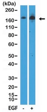
Evaluated by Western Blotting in lysate from A431 cells treated with EGF.

Western Blotting Analysis (WB): A 1:10,000 dilution of this antibody detected EGFR phosphorylated on Tyrosine 1068 in lysate from over-night serum starved A431 cells treated with EGF (50 ng/mL; 10 min.).

Tested Applications

Western Blotting Analysis: A 1:10,000 dilution from a representative lot detected Phospho-EGFR (Tyr1068) in lysate from HeLa cells overnight serum starved and treated with EGF ( (50 ng/mL; 10 min.).

Affinity Binding Assay: A representative lot of this antibody bound Phospho-EGF Receptor-(Tyr1068) peptide with a KD of 1.1 x 10⁻⁶ in an affinity binding assay.

Flow Cytometry Analysis: 1 µg from a representative lot detected Phospho-EGFR (Tyr1068) in one million A431 cells treated with EGF (100 ng/mL; 15 min.).

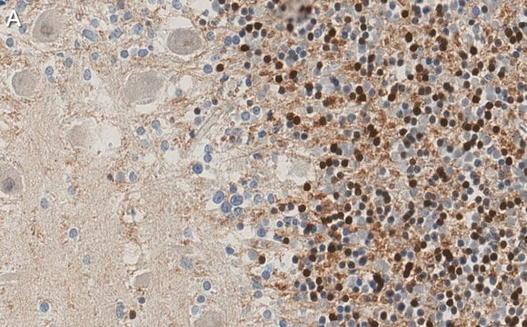
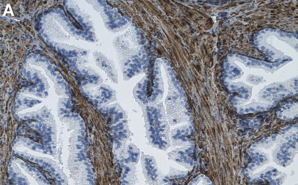
Immunohistochemistry (Paraffin) Analysis: A 1:100 dilution from a representative lot detected Phospho-EGFR (Tyr1068) in human cervical cancer tissue sections.

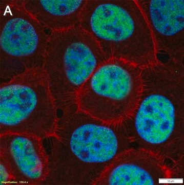
Immunocytochemistry Analysis: A 1:100 dilution from a representative lot detected Phospho-EGFR (Tyr1068) in A431 cells treated with EGF (200 ng/mL; 10 min.).

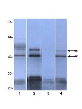
Peptide Inhibition Assay: Target band detection in a lysate from A431 cells treated with EGF (50 ng/mL; 10 min) was prevented by preblocking of a representative lot with the immunogen phosphopeptide, but not the corresponding non-phosphopeptide.

Note: Actual optimal working dilutions must be determined by end user as specimens, and experimental conditions may vary with the end user.

Target description

Epidermal growth factor receptor (UniProt: P00533; also known as EC:2.7.10.1, Proto-oncogene c-ErbB-1, Receptor tyrosine-protein kinase erbB-1) is encoded by the EGFR (also known as ERBB, ERBB1, HER1) gene (Gene ID: 1956) in human. EGFR is a single-pass type I membrane glycoprotein that is ubiquitously expressed. It is synthesized with a signal peptide (1-24), which is subsequently cleaved off to produce the mature form that contains an extracellular domain (aa 25-645), a transmembrane domain (aa 646-668), and a cytoplasmic domain (669-1210). It possesses receptor tyrosine kinase activity and upon binding of ligands activates several signaling cascades. Ligand binding is known to trigger receptor homo- and/or heterodimerization, which stimulates its intrinsic intracellular protein-tyrosine kinase activity. As a result, autophosphorylation of tyrosine residues in the C-terminal domain of EGFR occurs, which results in activation of several downstream signaling cascades, including the Ras-RAF-MEK-ERK, PI3 kinase-Akt, PLCγ-PKC, and STAT. EGFR phosphorylated on at serine 695 is reported to be only partial and occurs only if threonine 693 is phosphorylated.  Tyrosine 1068 is one of the major autophosphorylation sites. Phosphorylation at tyrosine 1068 is considered as a powerful biomarker associated with erlotinib sensitivity in vitro and in preclinical cancer stem cells generated xenografts. EGFR also undergoes mono and polyubiquitination upon EGF stimulation. However, this ubiquitination does not affect tyrosine kinase activity of the receptor or its signaling capacity. EGFR is commonly overexpressed and is mutated in many human malignancies and is often associated aggressive phenotypes. This ZooMAb® recombinant monoclonal antibody, generated by our propriety technology, offers significantly enhanced specificity, affinity, reproducibility, and stability over conventional monoclonals. (Ref.: Sette, G., et al. (2015). Cell Death Dis. 6(8); e1850; Wang, F., et al. (2012). J. Exp. Clin. Cancer Res. 31; Article 65).

Physical form

Purified recombinant rabbit monoclonal antibody IgG, lyophilized in PBS, 5% Trehalose, normal appearance a coarse or translucent resin. The PBS/trehalose components in the ZooMAb formulation can have the appearance of a semi-solid (bead like gel) after lyophilization. This is a normal phenomenon. Please follow the recommended reconstitution procedure in the data sheet to dissolve the semi-solid, bead-like, gel-appearing material. The resulting antibody solution is completely stable and functional as proven by full functional testing. Contains no biocide or preservatives, such as azide, or any animal by-products. Larger pack sizes provided as multiples of 25 µL.

Reconstitution

300 µg/mL after reconstitution at 25 µL per vial. Please refer to guidance on suggested starting dilutions and/or titers per application and sample type.

Storage and Stability

Recommend storage of lyophilized product at 2-8°C; Before reconstitution, micro-centrifuge vials briefly to spin down material to bottom of the vial; Reconstitute each vial by adding 25 µL of filtered lab grade water or PBS; Reconstituted antibodies can be stored at 2-8°C, or -20°C for long term storage. Avoid repeated freeze-thaws.

Other Notes

Concentration: Please refer to the Certificate of Analysis for the lot-specific concentration.

Legal Information

ZooMAb is a registered trademark of Merck KGaA, Darmstadt, Germany

Disclaimer

Unless otherwise stated in our catalog or other company documentation accompanying the product(s), our products are intended for research use only and are not to be used for any other purpose, which includes but is not limited to, unauthorized commercial uses, in vitro diagnostic uses, ex vivo or in vivo therapeutic uses or any type of consumption or application to humans or animals.

Not finding the right product?  

Try our Product Selector Tool.

Storage Class Code

11 - Combustible Solids

WGK

WGK 1

Flash Point(F)

Not applicable

Flash Point(C)

Not applicable


Choose from one of the most recent versions:

Certificates of Analysis (COA)

Lot/Batch Number

Don't see the Right Version?

If you require a particular version, you can look up a specific certificate by the Lot or Batch number.

Already Own This Product?

Find documentation for the products that you have recently purchased in the Document Library.

Visit the Document Library

Articles

Learn about the hallmarks of cancer and find reliable ZooMAb® recombinant antibodies to study them, conveniently organized by cancer hallmark.

Our team of scientists has experience in all areas of research including Life Science, Material Science, Chemical Synthesis, Chromatography, Analytical and many others.

Contact Technical Service