Skip to Content
Merck
All Photos(4)

Key Documents

Safety Information

AB1543P

Sigma-Aldrich

Anti-Synapsin I Antibody

Chemicon®, from rabbit

Synonym(s):

Synapsin-1

Sign Into View Organizational & Contract Pricing


About This Item

UNSPSC Code:
12352203
eCl@ss:
32160702
NACRES:
NA.41

biological source

rabbit

Quality Level

antibody form

affinity purified immunoglobulin

antibody product type

primary antibodies

clone

polyclonal

species reactivity

human, mouse, rat, bovine

manufacturer/tradename

Chemicon®

technique(s)

ELISA: suitable
immunocytochemistry: suitable
immunohistochemistry (formalin-fixed, paraffin-embedded sections): suitable
immunoprecipitation (IP): suitable
western blot: suitable

NCBI accession no.

UniProt accession no.

shipped in

dry ice

target post-translational modification

unmodified

Gene Information

human ... SYN1(6853)

General description

The synapsins are a family of proteins that have long been implicated in the regulation of neurotransmitter release at synapses. Specifically, they are thought to be involved in regulating the number of synaptic vesicles available for release via exocytosis at any one time. Synapsins are encoded by three different genes, synapsin I, II and III, and different neuron terminals will encode different amounts of these; synapsin will make up 1% of total brain protein at any one time.
Current studies suggest the following hypothesis for the role of synapsin: synapsins bind synaptic vesicles to components of the cytoskeleton which prevents them from migrating to the presynaptic membrane and releasing transmitter. During an action potential, synapsins are phosphorylated by Ca2+/calmodulin-dependent protein kinase II, releasing the synaptic vesicles and allowing them to move to the membrane and release their neurotransmitter.

Specificity

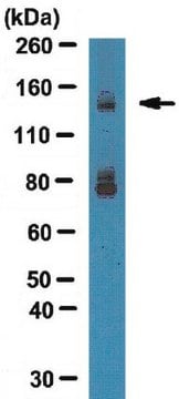
Synapsin I (Mr of 80,000 and 77,000). Synapsin is located only in brain nerve terminal and is an excellent marker for synapes. Immunolabeling is blocked by preadsorption of the antibody with synapsin I.

Immunogen

Synapsin I (mixture of Ia & Ib) purified from bovine brain.

Application

Research Category
Neuroscience
Research Sub Category
Synapse & Synaptic Biology
This Anti-Synapsin I Antibody is validated for use in ELISA, IC, IH, IH(P), IP, WB for the detection of Synapsin I.
Western blotting:
1:200-1:1,000 dilution of a previous lot was used.

Immunocytochemistry:
1:500-1:2,000 dilution of a previous lot was used.

Immunoprecipitation:
1 μg of a previous lot immunoprecipitated all of the synapsin I from a SDS homogenate of 200 μg of rat brain protein.

Note: The above dilutions are with 35S-protein A; with ECL dilutions may need to be considerably higher to obtain specific immunolabeling.

Immunohistochemistry:
1:500-1:2,500

ELISA:
1:2,500-1:10,00

Optimal working dilutions must be determined by the end user.

Quality

Immunohistochemistry(paraffin):
Synapsin I (AB1543P) representative staining pattern/morphology in rat hippocampal neurons. Tissue was pretreated with Citrate pH 6.0, antigen retrieval. This lot of antibody was diluted to 1:1000, IHC-Select reagents used with HRP-DAB. Immunoreactivity is seen directly associated with terminal end of neuronal cell body.
IHC-Paraffin Staining With Epitope Retrieval:
Rat Hippocampus

Target description

77 & 80 kDa

Linkage

Replaces: MAB355

Physical form

Format: Purified
ImmunoAffinity Purified
Purified rabbit polyclonal in buffer lyophilized from 5 mM ammonium bicarbonate. Reconstitute with 50 μL of PBS.

Storage and Stability

Stable for 1 year at -20ºC from date of receipt.

Analysis Note

Control
Brain tissue.

Other Notes

Concentration: Please refer to the Certificate of Analysis for the lot-specific concentration.

Legal Information

CHEMICON is a registered trademark of Merck KGaA, Darmstadt, Germany

Disclaimer

Unless otherwise stated in our catalog or other company documentation accompanying the product(s), our products are intended for research use only and are not to be used for any other purpose, which includes but is not limited to, unauthorized commercial uses, in vitro diagnostic uses, ex vivo or in vivo therapeutic uses or any type of consumption or application to humans or animals.

Not finding the right product?  

Try our Product Selector Tool.

Storage Class Code

11 - Combustible Solids

WGK

WGK 1


Regulatory Listings

Regulatory Listings are mainly provided for chemical products. Only limited information can be provided here for non-chemical products. No entry means none of the components are listed. It is the user’s obligation to ensure the safe and legal use of the product.

JAN Code

AB1543P:


Certificates of Analysis (COA)

Search for Certificates of Analysis (COA) by entering the products Lot/Batch Number. Lot and Batch Numbers can be found on a product’s label following the words ‘Lot’ or ‘Batch’.

Already Own This Product?

Find documentation for the products that you have recently purchased in the Document Library.

Visit the Document Library

Zi-Xuan He et al.
Cell reports, 33(5), 108343-108343 (2020-11-05)
Major depressive disorder (MDD) presents with two primary symptoms: depressed mood and anhedonia, which suggests that distinct neuronal circuits may regulate MDD. However, the underlying circuits of these individual symptoms linked to depression remain elusive. Herein, we identify a discrete
Diffuse and specific tectopulvinar terminals in the tree shrew: synapses, synapsins, and synaptic potentials.
Wei, H; Masterson, SP; Petry, HM; Bickford, ME
Testing null
Valerie Kilman et al.
The Journal of neuroscience : the official journal of the Society for Neuroscience, 22(4), 1328-1337 (2002-02-19)
Maintaining the proper balance between excitation and inhibition is necessary to prevent cortical circuits from either falling silent or generating epileptiform activity. One mechanism through which cortical networks maintain this balance is through the activity-dependent regulation of inhibition, but whether
NMDA receptors mediate synaptic competition in culture.
She, K; Craig, AM
Testing null
Katyayani Singh et al.
Brain research bulletin, 140, 5-18 (2018-04-02)
Cell surface neural adhesion proteins are critical components in the complex orchestration of cell proliferation, apoptosis, and neuritogenesis essential for proper brain construction and behavior. We focused on the impact of two plasticity-associated IgLON family neural adhesion molecules, Neurotrimin (Ntm)

Our team of scientists has experience in all areas of research including Life Science, Material Science, Chemical Synthesis, Chromatography, Analytical and many others.

Contact Technical Service