Skip to Content
Merck
All Photos(1)

Key Documents

Safety Information

AB15708A4

Sigma-Aldrich

Anti-Beta III Tubulin Antibody, Alexa Fluor 488 Conjugate | AB15708A4

from rabbit, ALEXA FLUOR 488

Synonym(s):

Tubulin beta-4, Tubulin beta-III, tubulin, beta 3, tubulin, beta, 4

Sign Into View Organizational & Contract Pricing


About This Item

UNSPSC Code:
12352203
eCl@ss:
32160702
NACRES:
NA.41

biological source

rabbit

Quality Level

conjugate

ALEXA FLUOR 488

antibody form

affinity isolated antibody

antibody product type

primary antibodies

clone

polyclonal

species reactivity

rat, mouse

species reactivity (predicted by homology)

human (based on 100% sequence homology)

technique(s)

immunocytochemistry: suitable
immunohistochemistry: suitable

NCBI accession no.

UniProt accession no.

shipped in

wet ice

target post-translational modification

unmodified

Gene Information

human ... TUBB3(10381)
mouse ... Tubb3(22152)
rat ... Tubb3(246118)

General description

Tubulin is one of several members of a small family of globular proteins which polymerize to form cytoskeletal structures called microtubules. The most common members of the tubulin family are α-tubulin and β-tubulin. Tubulin dimers, typically composed of α- and β-tubulin subunits, assemble onto microtubules forming large polymers. β-III tubulin is a unique microtubule subunit that is found expressed almost exclusively in neurons. Due to the unique expression pattern of β-III tubulin it is often utilized as a marker to identify neuronal cells from other brain cells such as glia which do not express it. β-III tubulin also serves as a cytoarchitectural marker since it is primarily found within neurite extensions.

Specificity

This antibody recognizes Beta III Tubulin.

Immunogen

Synthetic linear peptide corresponding to human Beta III Tubulin.

Application

Immunohistochemistry: Representive lot has been shown to work in adult mouse brain.
Research Category
Neuroscience
Research Sub Category
Neurofilament & Neuron Metabolism

Quality

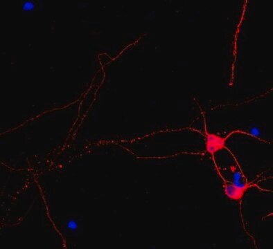
Evaluated by Immunocytochemistry in rat E18 cortex cells.

Immunocytochemistry Analysis: A 1:400 dilution of this antibody detected Beta III Tubulin in rat E18 cortex cells.

Target description

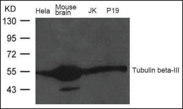
50 kDa calculated

Physical form

Affinity purified
Purified rabbit polyclonal conjugated to Alexa Fluor 488 in PBS with 0.1% sodium azide and 15 mg/mL BSA.

Storage and Stability

Stable for 1 year at 2-8°C from date of receipt.

Analysis Note

Control
Rat E18 cortex cells

Legal Information

ALEXA FLUOR is a trademark of Life Technologies

Disclaimer

Unless otherwise stated in our catalog or other company documentation accompanying the product(s), our products are intended for research use only and are not to be used for any other purpose, which includes but is not limited to, unauthorized commercial uses, in vitro diagnostic uses, ex vivo or in vivo therapeutic uses or any type of consumption or application to humans or animals.

Not finding the right product?  

Try our Product Selector Tool.

Storage Class Code

12 - Non Combustible Liquids

WGK

WGK 2

Flash Point(F)

Not applicable

Flash Point(C)

Not applicable


Regulatory Listings

Regulatory Listings are mainly provided for chemical products. Only limited information can be provided here for non-chemical products. No entry means none of the components are listed. It is the user’s obligation to ensure the safe and legal use of the product.

JAN Code

AB15708A4:


Certificates of Analysis (COA)

Search for Certificates of Analysis (COA) by entering the products Lot/Batch Number. Lot and Batch Numbers can be found on a product’s label following the words ‘Lot’ or ‘Batch’.

Already Own This Product?

Find documentation for the products that you have recently purchased in the Document Library.

Visit the Document Library

Predrag Kalaba et al.
Biomolecules, 13(9) (2023-09-28)
The high structural similarity, especially in transmembrane regions, of dopamine, norepinephrine, and serotonin transporters, as well as the lack of all crystal structures of human isoforms, make the specific targeting of individual transporters rather challenging. Ligand design itself is also
Jianzhang Hu et al.
Investigative ophthalmology & visual science, 60(1), 16-25 (2019-01-03)
To investigate the contribution and mechanism of miRNAs and autophagy in diabetic peripheral neuropathy. In this study, we used streptozotocin (STZ)-induced type I diabetes C57 mice as animal models, and we detected the expression of miR-34c and autophagic intensity in
Emily R Aurand et al.
Stem cell research, 12(1), 11-23 (2013-10-22)
Hydrogels provide a unique tool for neural tissue engineering. These materials can be customized for certain functions, i.e. to provide cell/drug delivery or act as a physical scaffold. Unfortunately, hydrogel complexities can negatively impact their biocompatibility, resulting in unintended consequences.
Zhenzhen Zhang et al.
Molecular vision, 24, 274-285 (2018-04-13)
To investigate the effect and mechanism of proresolving lipid mediator resolvin D1 (RvD1) on the corneal epithelium and the restoration of mechanical sensation in diabetic mice. Type 1 diabetes was induced in mice with intraperitoneal streptozocin injections. The healthy and
Yuan Zhang et al.
Frontiers in molecular biosciences, 8, 737472-737472 (2021-09-14)
Diabetes mellitus (DM) is a complex metabolic disorder. Long-term hyperglycemia may induce diabetic keratopathy (DK), which is mainly characterized by delayed corneal epithelial regeneration. MicroRNAs (miRNAs) have been reported to play regulatory roles during tissue regeneration. However, the molecular mechanism

Our team of scientists has experience in all areas of research including Life Science, Material Science, Chemical Synthesis, Chromatography, Analytical and many others.

Contact Technical Service