Skip to Content
Merck
All Photos(2)

Key Documents

Safety Information

AB5407

Sigma-Aldrich

Anti-Opsin Antibody, blue

Chemicon®, from rabbit

Synonym(s):

Anti-BCP, Anti-BOP

Sign Into View Organizational & Contract Pricing


About This Item

UNSPSC Code:
12352203
eCl@ss:
32160702
NACRES:
NA.41

biological source

rabbit

Quality Level

antibody form

purified immunoglobulin

antibody product type

primary antibodies

clone

polyclonal

species reactivity

monkey, human, mouse

manufacturer/tradename

Chemicon®

technique(s)

immunohistochemistry: suitable (paraffin)

NCBI accession no.

UniProt accession no.

shipped in

wet ice

target post-translational modification

unmodified

Gene Information

human ... OPN1LW(5956)

General description

The full range of color discrimination in humans is based on the presence and function of three cone photoreceptor mechanisms. Each cone type possesses a photo-sensitive pigment-protein complex consisting of 11-cis retinal and a unique opsin protein, which gives sensitivity in the short (S cone, peak sensitivity about 420nm), middle (M cone, peak sensitivity about 530nm with polymorphism; Winderckx et al., 1993; Neitz & Neitz, 1998), and long (L cone, peak sensitivity about 560nm with polymorphism; Neitz & Jacobs, 1990) wavelengths of the light spectrum. All three opsins are transmembrane proteins with seven membrane-spanning regions. Genes for the three types of cone opsins and the rod photoreceptor rhodopsin gene seem to be homologous with varying amounts of conservation. Strongest conservation is between the middle (green) and long (red) wavelength sensitive pigments on the X chromosome, suggesting a relatively recent duplication/divergence event (Nathans, 1989; Nathans et al., 1992). The S cone (blue) opsin is located on chromosome 7 and seems to have stronger conservation with rhodopsin. Cone photoreceptor distribution in humans is dominated by the M and L cone pigments.

Specificity

Recognizes Opsin, blue.

Immunogen

Epitope: blue
Recombinant human blue opsin.

Application

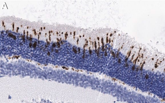
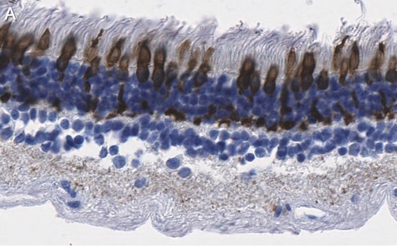
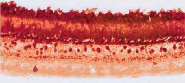
Immunohistochemistry: 1:200-1:300 on formalin-fixed, paraffin-embedded mouse retina tissue. Antigen retrieval method recommend is HIER with steam heat; other fixation and retrieval methods are untested.

Optimal working dilutions must be determined by the end user.
Research Category
Neuroscience
Research Sub Category
Sensory & PNS
This Anti-Opsin Antibody, blue is validated for use in IH(P) for the detection of Opsin.

Physical form

Format: Purified
Protein A purified
Purified immunoglobulinin PBS {0.02M phosphate, 0.25M NaCl, pH 7.6} with 0.1% sodium azide as a preservative

Storage and Stability

Maintain for 1 year at 2–8°C from date of shipment.

Analysis Note

Control
Retina

Legal Information

CHEMICON is a registered trademark of Merck KGaA, Darmstadt, Germany

Disclaimer

Unless otherwise stated in our catalog or other company documentation accompanying the product(s), our products are intended for research use only and are not to be used for any other purpose, which includes but is not limited to, unauthorized commercial uses, in vitro diagnostic uses, ex vivo or in vivo therapeutic uses or any type of consumption or application to humans or animals.

Not finding the right product?  

Try our Product Selector Tool.

Storage Class Code

12 - Non Combustible Liquids

WGK

WGK 1

Flash Point(F)

Not applicable

Flash Point(C)

Not applicable


Regulatory Listings

Regulatory Listings are mainly provided for chemical products. Only limited information can be provided here for non-chemical products. No entry means none of the components are listed. It is the user’s obligation to ensure the safe and legal use of the product.

JAN Code

AB5407:


Certificates of Analysis (COA)

Search for Certificates of Analysis (COA) by entering the products Lot/Batch Number. Lot and Batch Numbers can be found on a product’s label following the words ‘Lot’ or ‘Batch’.

Already Own This Product?

Find documentation for the products that you have recently purchased in the Document Library.

Visit the Document Library

Customers Also Viewed

Photoreceptor coupling mediated by connexin36 in the primate retina.
O'Brien, JJ; Chen, X; Macleish, PR; O'Brien, J; Massey, SC
The Journal of Neuroscience null
L P Morin et al.
Neuroscience, 199, 213-224 (2011-10-12)
Four studies were performed to further clarify the contribution of rod/cone and intrinsically photoreceptive retinal ganglion cells to measures of entrainment, dark preference, light-induced locomotor suppression and photosomnolence. Wild type (WT), retinally degenerate (rd/rd), and melanopsin-less (OPN4⁻/⁻) mouse strains were
Prasanthi Namburi et al.
American journal of human genetics, 99(3), 777-784 (2016-09-03)
Inherited retinal diseases (IRDs) are a diverse group of genetically and clinically heterogeneous retinal abnormalities. The present study was designed to identify genetic defects in individuals with an uncommon combination of autosomal recessive progressive cone-rod degeneration accompanied by sensorineural hearing
Histopathology and functional correlations in a patient with a mutation in RPE65, the gene for retinol isomerase.
Bonilha, VL; Rayborn, ME; Li, Y; Grossman, GH; Berson, EL; Hollyfield, JG
Investigative Ophthalmology & Visual Science null
Xiaojie Ji et al.
Investigative ophthalmology & visual science, 57(3), 877-888 (2016-03-16)
Retinal detachments (RDs), a separation of the light-sensitive tissue of the retina from its supporting layers in the posterior eye, isolate retinal cells from their normal supply of nourishment and can lead to their deterioration and death. We identified a

Our team of scientists has experience in all areas of research including Life Science, Material Science, Chemical Synthesis, Chromatography, Analytical and many others.

Contact Technical Service