Skip to Content
Merck
All Photos(2)

Key Documents

Safety Information

AB9864

Sigma-Aldrich

Anti-NMDAR1 Antibody, rabbit monoclonal

culture supernatant, clone 1.17.2.6, Chemicon®

Synonym(s):

N-methyl-D-aspartate receptor channel, subunit zeta-1, N-methyl-D-aspartate receptor subunit NR1, NMDA receptor 1, glutamate [NMDA] receptor subunit zeta 1, glutamate receptor, ionotropic, N-methyl D-aspartate 1

Sign Into View Organizational & Contract Pricing


About This Item

UNSPSC Code:
12352203
eCl@ss:
32160702
NACRES:
NA.41
clone:
1.17.2.6, monoclonal
application:
IHC
WB
species reactivity:
rat
technique(s):
immunohistochemistry: suitable
western blot: suitable
citations:
56

biological source

rabbit

Quality Level

antibody form

culture supernatant

antibody product type

primary antibodies

clone

1.17.2.6, monoclonal

species reactivity

rat

manufacturer/tradename

Chemicon®

technique(s)

immunohistochemistry: suitable
western blot: suitable

isotype

IgG

NCBI accession no.

UniProt accession no.

shipped in

wet ice

target post-translational modification

unmodified

Gene Information

General description

N-methyl-D-aspartate Receptors (NMDAR) are one of three pharmacologically distinct subtypes of ionotropic receptors that mediate a majority of excitatory neurotransmission in the brain via the endogenous amino acid, L-glutamate. MDARs form heteromeric complexes usually composed of two components, the NR1 subunit and NR2 subunits. Both proteins are weakly expressed throughout the brain but show localization in the dendritic region of the mushroom bodies. The NMDAR channel is highly permeable to Ca2+ and Na+ and its opening requires the simultaneous binding of glutamate and postsynaptic membrane depolarization. The activated NMDAR channel allows calcium influx into the post-synaptic cell where calcium triggers a cascade of biochemical events that create synaptic changes. NMDAR have been studied extensively in Drosophila for its potential relationship to behavioral plasticity. A newly identified NMDAR complex of more than 80 different proteins has been identified. Genetic and pharmacological disruptions of components of the NMDAR complex produce learning impairments in rodents, further linking behavioral plasticity to synaptic plasticity.

Specificity

Reactivity in mouse is weak. On reduced Western blots a band matching that in rat is observed but tests in immunohistochemistry have been unsuccessful. It is expected that the antibody will also react with monkey, gerbil, rabbit, feline and fish based on the fact that another antibody made to the same peptide sequence reacts with those species.
The original sequence used is selective for splice variants NR1-1a, NR1-1b, NR1-2a, NR1-2b using the nomenclature defined by Hollmann, M. et al. (1993). These appear to be the major splice variants expressed in rat brain (Sugihara H, et al., 1992). No cross-reaction with other glutamate receptor subunits. Western Blot analysis shows that this rabbit monoclonal antibody labels a single band at approximately 120 kD corresponding to NMDAR1 in rat brain lysate.

Immunogen

Epitope: a.a. 909-938
Synthetic peptide corresponding to the C-terminus of rat NMDA receptor subunit (Catalog number AG344). {amino acids 909-938 rat NMDAR1{LQNQKDTVLPRRAIERE EGQLQLCSRHRES}

Application

Immunohistochemistry:
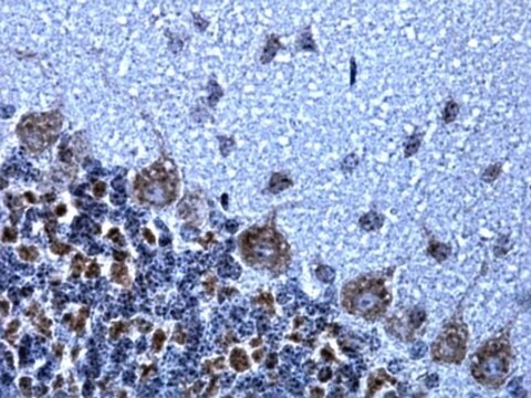
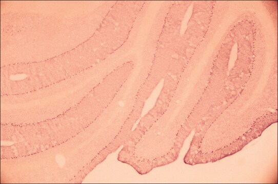
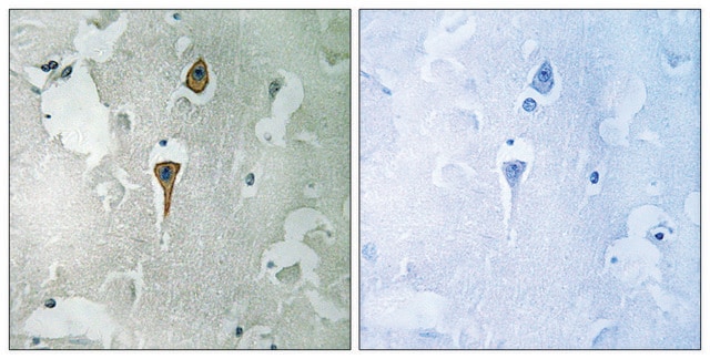
AB9864 can be used for using 4% paraformaldehyde or paraformaldehyde/glutaraldehyde fixed rat tissue. Suggested working dilution for light microscopy is 1:1,000-1:2,000.

Western blot:
1:1,000-1:2,000 using ECL on rat brain lysate.

Optimal working dilutions must be determined by end user.
Research Category
Neuroscience
Research Sub Category
Neurotransmitters & Receptors
This Anti-NMDAR1 Antibody is validated for use in IH, WB for the detection of NMDAR1.

Quality

Routinely evaluated by Western Blot on rat brain lysates.

Western Blot Analysis:
1:1000 dilution of this lot detected NMDAR1 on 10 μg of rat brain lysates

Target description

120 kDa

Linkage

Replaces: AB1516; 07-362

Physical form

Rabbit monoclonal supernatant liquid containing 0.05% sodium azide.
Unpurified

Storage and Stability

Stable for 1 year at -20ºC from date of receipt.

Analysis Note

Control
Brain tissue

Other Notes

Concentration: Please refer to the Certificate of Analysis for the lot-specific concentration.

Legal Information

CHEMICON is a registered trademark of Merck KGaA, Darmstadt, Germany

Disclaimer

Unless otherwise stated in our catalog or other company documentation accompanying the product(s), our products are intended for research use only and are not to be used for any other purpose, which includes but is not limited to, unauthorized commercial uses, in vitro diagnostic uses, ex vivo or in vivo therapeutic uses or any type of consumption or application to humans or animals.

Not finding the right product?  

Try our Product Selector Tool.

Storage Class Code

12 - Non Combustible Liquids

WGK

WGK 2


Regulatory Listings

Regulatory Listings are mainly provided for chemical products. Only limited information can be provided here for non-chemical products. No entry means none of the components are listed. It is the user’s obligation to ensure the safe and legal use of the product.

JAN Code

AB9864:


Certificates of Analysis (COA)

Search for Certificates of Analysis (COA) by entering the products Lot/Batch Number. Lot and Batch Numbers can be found on a product’s label following the words ‘Lot’ or ‘Batch’.

Already Own This Product?

Find documentation for the products that you have recently purchased in the Document Library.

Visit the Document Library

Brooke L Sinnen et al.
The Journal of neuroscience : the official journal of the Society for Neuroscience, 36(45), 11532-11543 (2016-12-03)
Beta amyloid (Aβ) triggers the elimination of excitatory synaptic connections in the CNS, an early manifestation of Alzheimer's disease. Oligomeric assemblies of Aβ peptide associate with excitatory synapses resulting in synapse elimination through a process that requires NMDA-type glutamate receptor
Tom J Phillips et al.
Scientific reports, 7(1), 9079-9079 (2017-08-24)
Some neuropsychiatric disease, including schizophrenia, may originate during prenatal development, following periods of gestational hypoxia and placental oxidative stress. Here we investigated if gestational hypoxia promotes damaging secretions from the placenta that affect fetal development and whether a mitochondria-targeted antioxidant
Gang Song et al.
Brain structure & function, 220(5), 2967-2982 (2014-07-25)
Current cellular-based connectomics approaches aim to delineate the functional or structural organizations of mammalian brain circuits through neuronal activity mapping and/or axonal tracing. To discern possible connectivity between functionally identified neurons in widely distributed brain circuits, reliable and efficient network-based
Catherine Croft Swanwick et al.
Developmental neurobiology, 70(13), 875-883 (2010-07-30)
Synapse malformation underlies numerous neurodevelopmental illnesses, including autism spectrum disorders. Here we identify the lipid raft protein flotillin-1 as a promoter of glutamatergic synapse formation. We cultured neurons from the hippocampus, a brain region important for learning and memory, and
Ling-Hsuan Kung et al.
PloS one, 8(7), e68312-e68312 (2013-07-12)
This study examines key elements of glutamatergic transmission within sensory ganglia of the rat. We show that the soma of primary sensory neurons release glutamate when depolarized. Using acute dissociated mixed neuronal/glia cultures of dorsal root ganglia (DRG) or trigeminal

Our team of scientists has experience in all areas of research including Life Science, Material Science, Chemical Synthesis, Chromatography, Analytical and many others.

Contact Technical Service