Skip to Content
Merck
All Photos(1)

Key Documents

Safety Information

ABT1373M

Sigma-Aldrich

Anti-Aggrecan

serum, from rabbit

Synonym(s):

Aggrecan core protein, Cartilage-specific proteoglycan core protein, CSPCP

Sign Into View Organizational & Contract Pricing


About This Item

UNSPSC Code:
12352203
eCl@ss:
32160702
NACRES:
NA.43
clone:
polyclonal
application:
IHC (p)
RIA
WB
species reactivity:
rat
technique(s):
immunohistochemistry (formalin-fixed, paraffin-embedded sections): suitable
radioimmunoassay: suitable
western blot: suitable
citations:

biological source

rabbit

Quality Level

antibody form

serum

antibody product type

primary antibodies

clone

polyclonal

species reactivity

rat

packaging

antibody small pack of 25 μL

technique(s)

immunohistochemistry (formalin-fixed, paraffin-embedded sections): suitable
radioimmunoassay: suitable
western blot: suitable

isotype

IgG

NCBI accession no.

UniProt accession no.

target post-translational modification

unmodified

Gene Information

rat ... Ncan(58982)

General description

Aggrecan core protein (UniProt: P07897; also known as Cartilage-specific proteoglycan core protein, CSPCP) is encoded by the Acan (also known as Agc, Agc1) gene (GeneID: 58968) in rat. Aggrecan is a secreted proteoglycan that is a major component of extracellular matrix of cartilagenous tissues. Its major function is to resist compression in cartilage. It binds avidly to hyaluronic acid via an N-terminal globular region and may play a regulatory role in the matrix assembly of the cartilage. Aggrecan contains two globular domains, G1 and G2 that comprise the N-terminus of the proteoglycan, while another globular region, G3, makes up the C-terminus. The G1 region is located in the N-terminus of the core protein and interacts with hyaluronic acid (HA). G1 contains Link domains and thus consists of three disulfide-bonded loop structures designated as the A, B, B′ motifs. The G2 region is composed of two B-like domains, but does not have the ability to interact with HA. G2 region is separated from the G1 region by the interglobular domain, which is a prominent site for proteolysis. The keratan sulfate (KS) and the chondroitin sulfate (CS) attachment domains lie between G2 and G3. Aggrecan is present in the extracellular matrix in the form of proteoglycan aggregates, in which many aggrecan molecules interact with hyaluronan and a link protein stabilizes each interaction. It is reported that aggrecan structure changes throughout the life cycle due to changes in synthetic and degradative events. Changes due to synthesis alter the structure of the chondroitin sulfate and keratan sulfate chains, whereas those due to degradation cause cleavage of all components of the aggregate. Aggrecan is also present in most of the retina including the outer nuclear layer, the outer plexiform layer, and the inner plexiform layer.

Specificity

This rabbit polyclonal antibody detects aggrecan in rat eye sections.

Immunogen

Epitope: unknown
GST-tagged recombinant fragment corresponding to 213 amino acids from the C-terminal half of rat Aggrecan.

Application

Anti-Aggrecan, Cat. No. ABT1373, is a rabbit polyclonal antibody that detects Aggrecan core protein and is tested for use in Immunohsitochemistry, Immunohistochemistry (Paraffin), Radioimmunoassay, and Western Blotting.
Radioimmunoassay Analysis: A representative lot detected Aggrecan in Postnatal rat brain (Milev, P., et. al. (1998). Biochem Biopys Res Commun. 247(2):207-12).

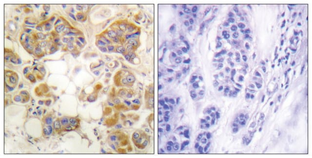
Immunohistochemistry Analysis: A representative lot detected Aggrecan in Embryonic day 16 rat eye, P0, P9 and adult rat retina (Popp, S., et. al. (2004). Exp Eye Res. 79(3):351-6) and Embryonic day 16, 19 rat brain and postnatal day 7 cerebellum, embryonic day 19 spinal cord (Popp, S., et. al. (2003). Dev Dyn. 227(1):143-9).

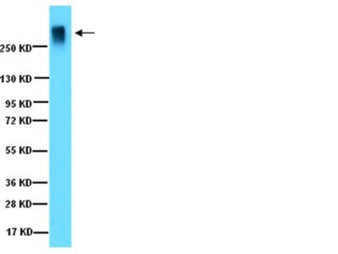
Western Blotting Analysis: A representative lot detected Aggrecan in purified proteoglycans from a PBS extract of 7-day rat brain with chondroitinase ABC treatment (Popp, S., et. al. (2003). Dev Dyn. 227(1):143-9).
Research Category
Cell Structure

Quality

Evaluated by Immunohistochemistry in rat retina tissue.

Immunohistochemistry Analysis: A 1:250 dilution of this antibody detected Aggrecan in rat retina tissue.

Target description

221.12 kDa calculated

Physical form

Rabbit polyclonal antiserum with 0.05% sodium azide.
Unpurified

Storage and Stability

Stable for 1 year at -20°C from date of receipt.

Other Notes

Concentration: Please refer to lot specific datasheet.

Disclaimer

Unless otherwise stated in our catalog or other company documentation accompanying the product(s), our products are intended for research use only and are not to be used for any other purpose, which includes but is not limited to, unauthorized commercial uses, in vitro diagnostic uses, ex vivo or in vivo therapeutic uses or any type of consumption or application to humans or animals.

Not finding the right product?  

Try our Product Selector Tool.

Storage Class Code

10 - Combustible liquids

WGK

WGK 1


Regulatory Listings

Regulatory Listings are mainly provided for chemical products. Only limited information can be provided here for non-chemical products. No entry means none of the components are listed. It is the user’s obligation to ensure the safe and legal use of the product.

JAN Code

ABT1373:
ABT1373-25UL:


Certificates of Analysis (COA)

Search for Certificates of Analysis (COA) by entering the products Lot/Batch Number. Lot and Batch Numbers can be found on a product’s label following the words ‘Lot’ or ‘Batch’.

Already Own This Product?

Find documentation for the products that you have recently purchased in the Document Library.

Visit the Document Library

Our team of scientists has experience in all areas of research including Life Science, Material Science, Chemical Synthesis, Chromatography, Analytical and many others.

Contact Technical Service