Skip to Content
Merck
All Photos(1)

Key Documents

Safety Information

CBL1337-I

Sigma-Aldrich

Anti-PECAM-1 Antibody, clone 390

clone 390, from rat

Synonym(s):

Platelet endothelial cell adhesion molecule, CD31, PECAM-1

Sign Into View Organizational & Contract Pricing


About This Item

UNSPSC Code:
12352203
eCl@ss:
32160702

biological source

rat

Quality Level

antibody form

purified immunoglobulin

antibody product type

primary antibodies

clone

390, monoclonal

species reactivity

mouse

technique(s)

flow cytometry: suitable
immunofluorescence: suitable
immunoprecipitation (IP): suitable
neutralization: suitable

isotype

IgG2aκ

NCBI accession no.

UniProt accession no.

shipped in

ambient

target post-translational modification

unmodified

Gene Information

mouse ... Pecam1(18613)

Related Categories

General description

Platelet endothelial cell adhesion molecule (UniProt Q08481; also known as CD31, PECAM-1) is encoded by the Pecam1 (also known as Pecam, Pecam-1) gene (Gene ID 18613) in murine species. PECAM-1 (CD31) is a 130-kD cell adhesion molecule that is expressed at the junctions of all continuous endothelia, as well as most bone marrow-derived hematopoietic cells, including platelets, monocytes, granulocytes and some lymphocytes. The human PECAM1 and murine Pecam1 genes both produce multiple spliced isoforms with varying lengths in cytoplasmic domain, with the isoform Long being the major form expressed in human tissues and endothelial cells (ECs), and isoform delta 14-15 being the predominantly expressed murine form. PECAM-1 plays an import role in maintaining vascular integrity and function by mediating ECs interactions with pericytes and smooth muscle cells (SMCs). PECAM-1 is initially produced with a signal peptide (human a.a. 1-27, murine a.a. 1-17) sequence, the removal of which yields the mature protein with a large extracellular region (human a.a. 28-601, mouse a.a. 18-590) composed of six C2-type Ig-like domains, followed by a transmembrane seqment (human a.a. 602-620, mouse a.a. 591-609) and a cytoplasmic tail (human a.a. 621-738, mouse a.a. 610-727) that plays an essential role in PECAM-1-mediated cellular signaling.

Specificity

Clone 390 immunoprecipitated the characteristic 120-130 kDa protein from L-cells transfected with muPECAM-1∆12,15 and selectively stained murine vessels in acetone-fixed tissue sections (Baldwin, H.S., et al. (1994). Development. 120(9):2539-2553).

Immunogen

Epitope: extracellular domain
Mouse leukocyte cell line 32D.

Application

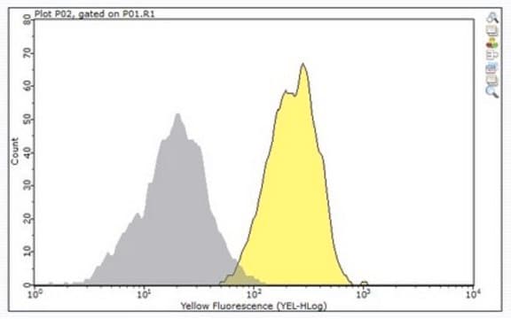
Flow Cytometry Analysis: A representative lot detected the expression of wild-type, ∆12,15 and ∆14,15 murine PECAM-1 constructs on the surface of transfected mouse L-cells (Baldwin, H.S., et al. (1994). Development. 120(9):2539-2553).

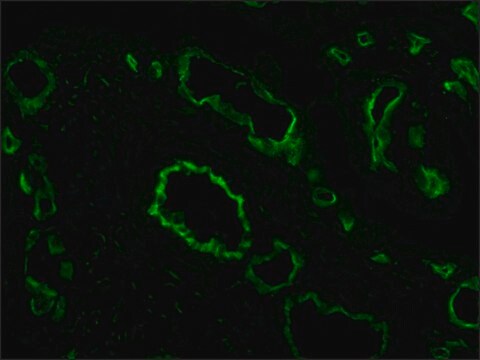
Immunofluorescence Analysis: A representative lot localized PECAM-1 immunoreactivity by fluorescent immunohistochemistry staining of murine embryo cryosections (Baldwin, H.S., et al. (1994). Development. 120(9):2539-2553).

Immunoprecipitation Analysis: A representative lot immunoprecipitated the 120-130 kDa exogenously expressed PECAM-1 ∆12,15 construct from transfected murine L-cells (Baldwin, H.S., et al. (1994). Development. 120(9):2539-2553).

Neutralizing Analysis: A representative lot inhibited the aggregation of murine L-cell transfectants expressing wild-type and ∆12,15, but not ∆14,15, murine PECAM-1 constructs (Baldwin, H.S., et al. (1994). Development. 120(9):2539-2553).

Clone 390 is also available as FITC conjugate (Cat. No. CBL1337F) for direct immunofluorescent staining in flow cytometry, immunocytochemistry and immunohistochemistry applications.
Research Category
Cell Structure
This rat monoclonal Anti-PECAM-1 Antibody, clone 390, Cat. No. CBL1337-I is validated for use in Flow Cytometry, Immunofluorescence, Immunoprecipitation, and Neutralization applications.

Quality

Evaluated by Flow Cytometry in mouse splenocytes.

Flow Cytometry Analysis: 2 µL of this antibody detected PECAM-1-positive monocytes among one million mouse splenocytes.

Target description

79.49/76.14/78.23/68.05 kDa (mature isoform 1/2/3/4), 81.26/77.92/80.00/69.82 kDa (isoform 1/2/3/4 pro-form) calculated.

Linkage

Replaces: CBL1337

Physical form

Format: Purified
Protein G purified.
Purified rat IgG2a in PBS without azide.

Storage and Stability

Stable for 1 year at -20°C from date of receipt.
Handling Recommendations: Upon receipt and prior to removing the cap, centrifuge the vial and gently mix the solution. Aliquot into microcentrifuge tubes and store at -20°C. Avoid repeated freeze/thaw cycles, which may damage IgG and affect product performance.

Other Notes

Concentration: Please refer to lot specific datasheet.

Disclaimer

Unless otherwise stated in our catalog or other company documentation accompanying the product(s), our products are intended for research use only and are not to be used for any other purpose, which includes but is not limited to, unauthorized commercial uses, in vitro diagnostic uses, ex vivo or in vivo therapeutic uses or any type of consumption or application to humans or animals.

Not finding the right product?  

Try our Product Selector Tool.

Storage Class Code

12 - Non Combustible Liquids

WGK

WGK 2

Flash Point(F)

Not applicable

Flash Point(C)

Not applicable


Regulatory Listings

Regulatory Listings are mainly provided for chemical products. Only limited information can be provided here for non-chemical products. No entry means none of the components are listed. It is the user’s obligation to ensure the safe and legal use of the product.

JAN Code

CBL1337-I:


Certificates of Analysis (COA)

Search for Certificates of Analysis (COA) by entering the products Lot/Batch Number. Lot and Batch Numbers can be found on a product’s label following the words ‘Lot’ or ‘Batch’.

Already Own This Product?

Find documentation for the products that you have recently purchased in the Document Library.

Visit the Document Library

Rose Z Hill et al.
Nature, 607(7917), 104-110 (2022-06-23)
Itch triggers scratching, a behavioural defence mechanism that aids in the removal of harmful irritants and parasites1. Chemical itch is triggered by many endogenous and exogenous cues, such as pro-inflammatory histamine, which is released during an allergic reaction1. Mechanical itch

Our team of scientists has experience in all areas of research including Life Science, Material Science, Chemical Synthesis, Chromatography, Analytical and many others.

Contact Technical Service