Skip to Content
Merck
All Photos(3)

Key Documents

Safety Information

MAB2079-AF647

Sigma-Aldrich

Anti-Integrin β1, activated, clone HUTS-4, Alexa Fluor 647 Conjugate

clone HUTS-4, from mouse, ALEXA FLUOR 647

Synonym(s):

Integrin beta-1, Fibronectin receptor subunit beta, Glycoprotein IIa, GPIIA, VLA-4 subunit beta, CD29, Integrin β1, activated

Sign Into View Organizational & Contract Pricing


About This Item

UNSPSC Code:
12352203
eCl@ss:
32160702
NACRES:
NA.41

biological source

mouse

Quality Level

conjugate

ALEXA FLUOR 647

antibody form

purified antibody

antibody product type

primary antibodies

clone

HUTS-4, monoclonal

species reactivity

mouse, human

species reactivity (predicted by homology)

rat (based on 100% sequence homology)

technique(s)

immunocytochemistry: suitable

isotype

IgG2a

NCBI accession no.

UniProt accession no.

shipped in

wet ice

target post-translational modification

unmodified

Gene Information

human ... ITGB1(3688)

General description

Integrin beta-1 (UniProt P05556; also known as CD29, Glycoprotein Iia, GPIIA, Integrin VLA-4 subunit beta, Very late activation protein, beta polypeptide) is encoded by the ITGB1 (also known as FNRB, MDF2, MSK12, VLA-BETA, VLAB) gene (Gene ID 3688) in human. Integrin beta-1 is a 130 kDa transmembrane glycoprotein that interacts with various integrin alpha subunits (including alpha 1, 2, 3, 4, 5 and 6) to form the functional receptor complexes that bind to specific extracellular matrix proteins. Integrin receptors regulate a variety of important biological functions, including embryonic development, wound repair, hemostasis, and prevention of programmed cell death. They are also implicated in abnormal pathological states such as tumor directed angiogenesis, tumor growth and metastasis. These heterodimeric receptors bridge the cytoplasmic actin cytoskeleton with proteins present in the extracellular matrix and/or on adjacent cells. Interactions between integrins and the extracellular matrix lead to the activation of signal transduction pathways and regulation of gene expression.

Specificity

Monoclonal antibody HUTS-4 is specific for active conformation of human beta1 integrins. The beta1 or VLA integrin subgroup contains at least 10 members, each with a distinct alpha subunit nonconvalently associated with the common beta1 subunit. Functional activation of integrins can be experimentally induced by addition of Mg2+ or Mn2+, or by using one of several stimulatory antibodies that are available. Intracellular signals generated as a result of cell activation also modulate the confirmation and affinity of integrins in cellular systems. Antibodies such as HUTS-4 which have the ability to discriminate between different states of integrin activation are exceptionally useful for studies on the regulation of activation. Antibody HUTS-4 recognizes epitopes in the 355-425 region of the common beta1 subunit of VLA integrins, whose expression parallels the ligand binding activity of these adhesion receptors induced by various extracellular and intracellular stimuli.

Immunogen

Epitope: A.a. 355-425 region of the common β1 subunit of VLA integrins.
Generated against human beta1 integrins purified from Triton X-100 lysates of human lung, liver, and skeletal muscle tissues.

Application

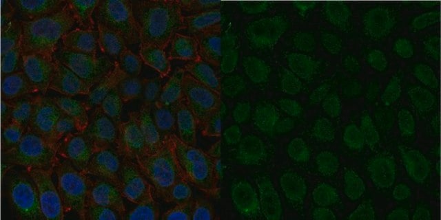
Immunocytochemistry Analysis: A 1:100 dilution from a representative lot detected Integrin β1 in HeLa and A431 cells.
The unconjugated antibody (Cat. No. MAB2079Z) is shown to be suitable also for Western blotting, ELISA, functional assay, and immunohistochemistry.
Research Category
Cell Structure
Research Sub Category
Adhesion (CAMs)
This Anti-Integrin β1, activated, clone HUTS-4, AlexaFluor 647 Conjugate is validated for use in Immunocytochemistry for the detection of Integrin β1.

Quality

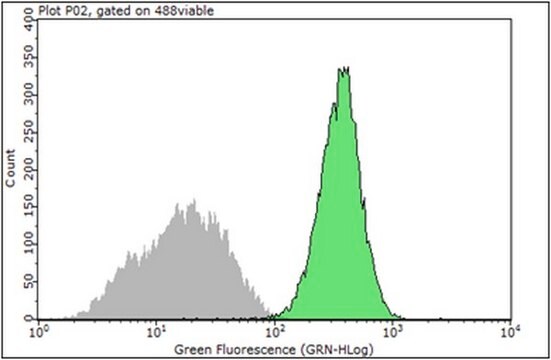
Evaluated by Immunocytochemistry in NIH/3T3 cells.

Immunocytochemistry Analysis: A 1:100 dilution of this antibody detected Integrin β1 in NIH/3T3 cells.

Target description

~88 kDa observed

Physical form

Protein G Purified
Purified mouse antibody conjugate in PBS with 15 mg/ml BSA and 0.1% sodium azide.

Storage and Stability

Stable for 1 year at 2-8°C from date of receipt.

Other Notes

Concentration: Please refer to lot specific datasheet.

Legal Information

ALEXA FLUOR is a trademark of Life Technologies

Disclaimer

Unless otherwise stated in our catalog or other company documentation accompanying the product(s), our products are intended for research use only and are not to be used for any other purpose, which includes but is not limited to, unauthorized commercial uses, in vitro diagnostic uses, ex vivo or in vivo therapeutic uses or any type of consumption or application to humans or animals.

Not finding the right product?  

Try our Product Selector Tool.

Storage Class Code

12 - Non Combustible Liquids

WGK

WGK 2

Flash Point(F)

Not applicable

Flash Point(C)

Not applicable


Regulatory Listings

Regulatory Listings are mainly provided for chemical products. Only limited information can be provided here for non-chemical products. No entry means none of the components are listed. It is the user’s obligation to ensure the safe and legal use of the product.

JAN Code

MAB2079-AF647:


Certificates of Analysis (COA)

Search for Certificates of Analysis (COA) by entering the products Lot/Batch Number. Lot and Batch Numbers can be found on a product’s label following the words ‘Lot’ or ‘Batch’.

Already Own This Product?

Find documentation for the products that you have recently purchased in the Document Library.

Visit the Document Library

Ayan T Ray et al.
Development (Cambridge, England), 150(6) (2023-03-03)
Loss of FGF signaling leads to defects in salivary gland branching, but the mechanisms underlying this phenotype remain largely unknown. We disrupted expression of Fgfr1 and Fgfr2 in salivary gland epithelial cells and found that both receptors function coordinately in
Marta Mastrogiovanni et al.
Science advances, 8(15), eabl5942-eabl5942 (2022-04-14)
Adenomatous polyposis coli (APC) is a tumor suppressor whose mutations underlie familial adenomatous polyposis (FAP) and colorectal cancer. Although its role in intestinal epithelial cells is well characterized, APC importance in T cell biology is ill defined. APC regulates cytoskeleton

Our team of scientists has experience in all areas of research including Life Science, Material Science, Chemical Synthesis, Chromatography, Analytical and many others.

Contact Technical Service