MAB5278
Anti-Tryptophan Hydroxylase/Tyrosine Hydroxylase/Phenylalanine Hydroxylase Antibody
CHEMICON®, mouse monoclonal, PH8
About This Item
Recommended Products
product name
Anti-Tryptophan Hydroxylase/Tyrosine Hydroxylase/Phenylalanine Hydroxylase Antibody, clone PH8, clone PH8, Chemicon®, from mouse
biological source
mouse
Quality Level
antibody form
purified immunoglobulin
antibody product type
primary antibodies
clone
PH8, monoclonal
species reactivity
human
manufacturer/tradename
Chemicon®
technique(s)
immunohistochemistry: suitable
immunoprecipitation (IP): suitable
western blot: suitable
isotype
IgG1
NCBI accession no.
shipped in
wet ice
target post-translational modification
unmodified
Gene Information
human ... TPH1(7166)
General description
Application
PROTOCOLS
Immunohistochemistry
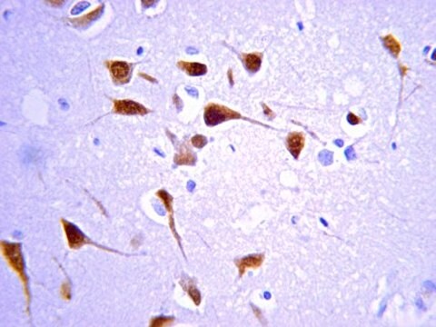
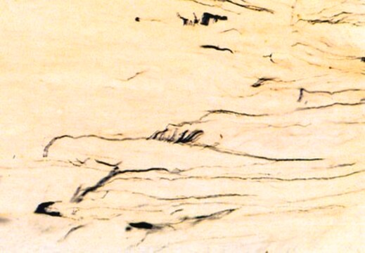
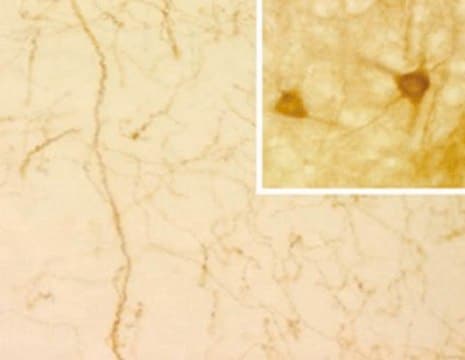
The PH8 Antibody can be used for the immunohistochemical detection of dopaminergic and serotonergic neurons in human and rat brain stem tissue.
1. Tissue should be formalin fixed and stored in formalin prior to use for a minimum of five days.
2. Cryoprotect tissue using 30% sucrose in 0.1M Tris pH 7.4 buffer for 24-72 hours.
3. Cut using a sledge microtome to 50 μm thickness.
4. Wash the tissue samples using Tris buffer prior to commencing the staining procedure.
5. Treat the tissue samples for 3 x 15 minutes in 50% alcohol.
6. Treat the tissue samples for 20 minutes in 50% alcohol and 3% H2O2.
7. Treat tissue for 20 minutes with 10% normal horse serum in Tris buffer. This acts to block endogeneous H2O2 staining.
8. Dilute the PH8 Antibody in Tris buffer, add to the tissue samples and incubate for 1-3 days. Recommended dilutions are 1:2,000-1:10,000. At high antibody concentration all monoaminergic neurons are stained with no distinction between serotonegic and catecholominergic cells. By diluting the antibody concentrate, cell types are distinguishable due to variation in staining intensity. At lower concentrations (1:5,000-1:10,000 dilution) only serotonergic cells will stain.
9. Wash tissue (3 x 15 minutes), then add a biotinylated anti-mouse secondary antibody and incubate on an orbital shaker at room temperature (RT) for 1 hour.
10. Wash tissue (3 x 15 minutes) and incubate on an orbital shaker at RT for 1 hour with the tertiary complex (ELITE KIT, Vector, USA).
11. Wash tissue (3 x 15 minutes) and incubate on an orbital shaker at RT for 10 minutes with Tris buffered diamino-benzidine substrate. Add 0.1% H2O2 and incubate on an orbital shaker at RT for a further 5 minutes.
12. Mount tissue onto gelatinised slides and allow to dry prior to microscopy.
Physical form
Storage and Stability
Legal Information
Not finding the right product?
Try our Product Selector Tool.
Storage Class Code
12 - Non Combustible Liquids
WGK
WGK 2
Flash Point(F)
Not applicable
Flash Point(C)
Not applicable
Regulatory Listings
Regulatory Listings are mainly provided for chemical products. Only limited information can be provided here for non-chemical products. No entry means none of the components are listed. It is the user’s obligation to ensure the safe and legal use of the product.
JAN Code
MAB5278:
Certificates of Analysis (COA)
Search for Certificates of Analysis (COA) by entering the products Lot/Batch Number. Lot and Batch Numbers can be found on a product’s label following the words ‘Lot’ or ‘Batch’.
Already Own This Product?
Find documentation for the products that you have recently purchased in the Document Library.
Our team of scientists has experience in all areas of research including Life Science, Material Science, Chemical Synthesis, Chromatography, Analytical and many others.
Contact Technical Service


