Skip to Content
Merck
All Photos(1)

Key Documents

Safety Information

MAB5352

Sigma-Aldrich

Anti-Notch 1 Antibody, clone mN1A

clone mN1A, Chemicon®, from mouse

Synonym(s):

Anti-AOS5, Anti-AOVD1, Anti-TAN1, Anti-hN1

Sign Into View Organizational & Contract Pricing


About This Item

UNSPSC Code:
12352203
eCl@ss:
32160702
NACRES:
NA.41

biological source

mouse

Quality Level

antibody form

purified immunoglobulin

antibody product type

primary antibodies

clone

mN1A, monoclonal

species reactivity

rat, mouse, feline

packaging

antibody small pack of 25 μg

manufacturer/tradename

Chemicon®

technique(s)

immunocytochemistry: suitable
immunohistochemistry: suitable
immunoprecipitation (IP): suitable
western blot: suitable

isotype

IgG1

NCBI accession no.

UniProt accession no.

shipped in

ambient

storage temp.

2-8°C

target post-translational modification

unmodified

Gene Information

human ... NOTCH1(4851)

General description

Notch1 consists of three domains. The extracellular domain containing 36 epidermal growth factor (EGF) repeats and 3 lin-12/Notch repeats (LNR) and functions as a receptor in cell-cell interactions. There is a single transmembrane domain (TM). The intracellular domain contains six cdc10/ankyrin repeats (cdc10) and the newly defined Notch cytokine response (NCR) region, which contains a putative bipartite NLS (nuclear localization signal).

Specificity

Recognizes Notch1, intracellular domain. The antibody has a high affinity for the "activated" intracellular form of Notch1, but a low affinity for the full-length (unprocessed or heterodimeric cell surface) forms. No reactivity with Notch2, 3 or 4.

Immunogen

Cdc10-NCR region of mouse Notch1 expressed as a GST fusion protein.

Application

Anti-Notch 1 Antibody, clone mN1A is a high quality Mouse Monoclonal Antibody for the detection of Notch 1 & has been validated in ICC, IHC, IP & WB.
Research Category
Epigenetics & Nuclear Function
Research Sub Category
Transcription Factors
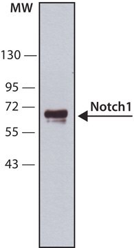
Western blot using ECL

Immunohistochemistry

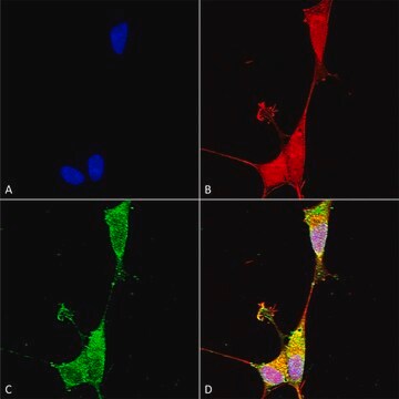
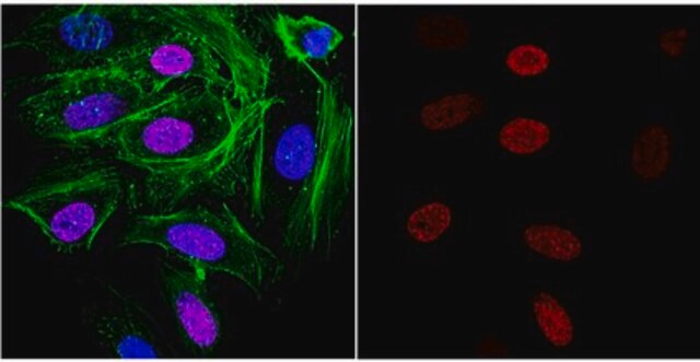
Immunocytochemistry

Immunoprecipitation

Optimal working dilutions must be determined by the end user.

Target description

272.5 kDa (also 230 kDa processed form)

Linkage

Replaces: 04-1046

Physical form

Format: Purified
Protein A purified
Purified immunoglobulin. Liquid.

Storage and Stability

Maintain for 1 year at 2–8°C from date of shipment. Aliquot to avoid repeated freezing and thawing. For maximum recovery of product, centrifuge the original vial after thawing and prior to removing the cap.

Analysis Note

Control
In fetal tissues, use spleen, brain stem and lung. In most adult tissues, use lymphoid tissues.

Other Notes

Concentration: Please refer to the Certificate of Analysis for the lot-specific concentration.

Legal Information

CHEMICON is a registered trademark of Merck KGaA, Darmstadt, Germany

Disclaimer

Unless otherwise stated in our catalog or other company documentation accompanying the product(s), our products are intended for research use only and are not to be used for any other purpose, which includes but is not limited to, unauthorized commercial uses, in vitro diagnostic uses, ex vivo or in vivo therapeutic uses or any type of consumption or application to humans or animals.

Not finding the right product?  

Try our Product Selector Tool.

Storage Class Code

10 - Combustible liquids

WGK

WGK 2

Flash Point(F)

Not applicable

Flash Point(C)

Not applicable


Regulatory Listings

Regulatory Listings are mainly provided for chemical products. Only limited information can be provided here for non-chemical products. No entry means none of the components are listed. It is the user’s obligation to ensure the safe and legal use of the product.

JAN Code

MAB5352-25UG:
MAB5352:


Certificates of Analysis (COA)

Search for Certificates of Analysis (COA) by entering the products Lot/Batch Number. Lot and Batch Numbers can be found on a product’s label following the words ‘Lot’ or ‘Batch’.

Already Own This Product?

Find documentation for the products that you have recently purchased in the Document Library.

Visit the Document Library

Fei Guo et al.
Cell biology international, 36(11), 997-1004 (2012-08-08)
Angiogenesis takes place after brain ischaemia, and stroke-induced angiogenesis in ischaemic brain may be associated with improved neurological recovery. Bone MSCs (marrow stromal cells) transplantation can promote this vital angiogenesis in ischaemic zones, but the mechanisms by which MSCs promoting
Xue-Liang Zhou et al.
Journal of cellular physiology, 234(6), 8834-8845 (2018-10-15)
During myocardial infarction (MI), cardiac fibroblasts (CFs) transform into myofibroblast (CMT). This study aimed to investigate the crosstalk of Notch1 and transforming growth factor-β1 (TGF-β1)/Smad3 signaling in the regulation of CMT and myocardial fibrosis. Primary CFs were isolated from young
Shu Li et al.
Molecular medicine reports, 9(6), 2233-2238 (2014-03-20)
Brain arteriovenous malformation (bAVM) is currently one of the most common cerebral vascular diseases, which result in severe clinical outcomes. The Notch signaling pathway is involved in vasculogenesis and angiogenesis, as well as vascular remodeling and arteriovenous differentiation in multiple
Yan-Min Feng et al.
Cell cycle (Georgetown, Tex.), 13(5), 782-791 (2014-01-09)
A critical process of early oogenesis is the entry of mitotic oogonia into meiosis, a cell cycle switch regulated by a complex gene regulatory network. Although Notch pathway is involved in numerous important aspects of oogenesis in invertebrate species, whether
Susanne Pettersson et al.
The Biochemical journal, 450(3), 523-536 (2012-12-21)
The Notch receptor is necessary for modulating cell fate decisions throughout development, and aberrant activation of Notch signalling has been associated with many diseases, including tumorigenesis. The E3 ligase MDM2 (murine double minute 2) plays a role in regulating the

Our team of scientists has experience in all areas of research including Life Science, Material Science, Chemical Synthesis, Chromatography, Analytical and many others.

Contact Technical Service