Skip to Content
Merck
All Photos(1)

Key Documents

Safety Information

MABC544

Sigma-Aldrich

Anti-PLK-4 Antibody, clone 6H5

clone 6H5, from mouse

Synonym(s):

Serine/threonine-protein kinase PLK4, Polo-like kinase 4, PLK-4, Serine/threonine-protein kinase 18, Serine/threonine-protein kinase Sak

Sign Into View Organizational & Contract Pricing


About This Item

UNSPSC Code:
12352203
eCl@ss:
32160702
NACRES:
NA.41

biological source

mouse

Quality Level

antibody form

purified immunoglobulin

antibody product type

primary antibodies

clone

6H5, monoclonal

species reactivity

human

technique(s)

immunocytochemistry: suitable
immunoprecipitation (IP): suitable
western blot: suitable

isotype

IgG3κ

NCBI accession no.

UniProt accession no.

shipped in

wet ice

target post-translational modification

unmodified

Gene Information

human ... PLK4(10733)

General description

Serine/threonine-protein kinase PLK4 (PLK-4) is a member of the protein kinase superfamily and plays an essential role in centriole duplication. PLK-4 triggers procentriole formation on the surface of the parental centriole cylinder, leading to the recruitment of centriole biogenesis proteins. It may be involved in tumorigenesis since centrosome aberrations are frequently observed in tumors. PLK-4 may also be involved in trophoblast differentiation by phosphorylating HAND1, leading to the disruption between HAND1 and MDFIC and then activating HAND1.

Immunogen

Linear peptide corresponding to Human PLK-4.

Application

Detect Polo Like Kinase (PLK) using this mouse monoclonal antibody, Anti-PLK-4 Antibody, clone 6H5 validated for use in western blotting, ICC & IP.
Immunocytochemistry Analysis: A representative lot detected PLK-4 in U20S cell lysates transfected with control and Cep152 siRNA, but not U2OS cell lysates transfected with PLK4 siRNA. (Cizmecioglu, O. et al. (2010). J Cell Biol. 191(4):731-739).
Immunoprecipitation Analysis: A representative lot immunoprecipitated PLK-4 in HeLa cell lysates cotransfected with Flag-tagged Gcp6 and Myc-tagged Plk-4 (Bahtz, R. et al. (2012). J Cell Sci. 125 (Pt 2): 486-496).
Research Category
Apoptosis & Cancer
Research Sub Category
Apoptosis - Additional

Quality

Evaluated by Western Blotting in U2Os cell lysate.

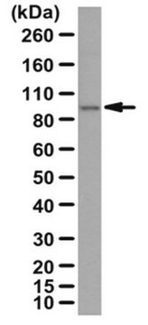
Western Blotting Analysis: 4.0 µg/mL of this antibody detected PLK-4 in 10 µg of U2Os cell lysate.

Target description

109 kDa observed

Physical form

Format: Purified
Protein G Purified
Purified mouse monoclonal IgG3κ in buffer containing 0.1 M Tris-Glycine (pH 7.4), 150 mM NaCl with 0.05% sodium azide.

Storage and Stability

Stable for 1 year at 2-8°C from date of receipt.

Analysis Note

Control
U2Os cell lysate

Other Notes

Concentration: Please refer to the Certificate of Analysis for the lot-specific concentration.

Disclaimer

Unless otherwise stated in our catalog or other company documentation accompanying the product(s), our products are intended for research use only and are not to be used for any other purpose, which includes but is not limited to, unauthorized commercial uses, in vitro diagnostic uses, ex vivo or in vivo therapeutic uses or any type of consumption or application to humans or animals.

Not finding the right product?  

Try our Product Selector Tool.

Storage Class Code

12 - Non Combustible Liquids

WGK

WGK 1

Flash Point(F)

Not applicable

Flash Point(C)

Not applicable


Regulatory Listings

Regulatory Listings are mainly provided for chemical products. Only limited information can be provided here for non-chemical products. No entry means none of the components are listed. It is the user’s obligation to ensure the safe and legal use of the product.

JAN Code

MABC544:


Certificates of Analysis (COA)

Search for Certificates of Analysis (COA) by entering the products Lot/Batch Number. Lot and Batch Numbers can be found on a product’s label following the words ‘Lot’ or ‘Batch’.

Already Own This Product?

Find documentation for the products that you have recently purchased in the Document Library.

Visit the Document Library

Midori Ohta et al.
Nature communications, 5, 5267-5267 (2014-10-25)
Formation of one procentriole next to each pre-existing centriole is essential for centrosome duplication, robust bipolar spindle assembly and maintenance of genome integrity. However, the mechanisms maintaining strict control over centriole copy number are incompletely understood. Here we show that
Onur Rojhat Karasu et al.
The Journal of cell biology, 221(12) (2022-11-01)
The centriole is the microtubule-based backbone that ensures integrity, function, and cell cycle-dependent duplication of centrosomes. Mostly unclear mechanisms control structural integrity of centrioles. Here, we show that the centrosome protein CEP350 functions as scaffold that coordinates distal-end properties of
Satoko Yoshiba et al.
Journal of cell science, 132(12) (2019-06-06)
At the onset of procentriole formation, a structure called the cartwheel is formed adjacent to the pre-existing centriole. SAS-6 proteins are thought to constitute the hub of the cartwheel structure. However, the exact function of the cartwheel in the process
Midori Ohta et al.
Cell reports, 23(11), 3160-3169 (2018-06-14)
The number of centrioles is tightly controlled to ensure bipolar spindle assembly, which is a prerequisite to maintain genome integrity. However, our understanding of the fundamental principle that governs the formation of a single procentriole per parental centriole is incomplete.
Dasom Gwon et al.
Molecules and cells, 42(12), 840-849 (2019-11-15)
The spatiotemporal mitotic processes are controlled qualitatively by phosphorylation and qualitatively by ubiquitination. Although the SKP1-CUL1-F-box protein (SCF) complex and the anaphase-promoting complex/cyclosome (APC/C) mainly mediate ubiquitin-dependent proteolysis of mitotic regulators, the E3 ligase for a large portion of mitotic

Our team of scientists has experience in all areas of research including Life Science, Material Science, Chemical Synthesis, Chromatography, Analytical and many others.

Contact Technical Service