Skip to Content
Merck
All Photos(2)

Key Documents

Safety Information

MABN11

Sigma-Aldrich

Anti-Amyloid β40 Antibody, clone G2-10

clone G2-10, from mouse

Synonym(s):

Alzheimer disease, Alzheimer disease amyloid protein, Cerebral vascular amyloid peptide, Protease nexin-II, amyloid beta (A4) precursor protein, amyloid beta A4 protein, amyloid beta precursor protein, beta-amyloid peptide, human mRNA for amyloid A4 prec

Sign Into View Organizational & Contract Pricing


About This Item

UNSPSC Code:
12352203
eCl@ss:
32160702
NACRES:
NA.41
clone:
G2-10, monoclonal
application:
ELISA
IHC
WB
species reactivity:
human, mouse
technique(s):
ELISA: suitable
immunohistochemistry: suitable
western blot: suitable
citations:
5

biological source

mouse

Quality Level

antibody form

purified antibody

clone

G2-10, monoclonal

species reactivity

human, mouse

technique(s)

ELISA: suitable
immunohistochemistry: suitable
western blot: suitable

isotype

IgG2bκ

NCBI accession no.

UniProt accession no.

shipped in

wet ice

target post-translational modification

unmodified

Gene Information

human ... APP(351)

General description

The cerebral and vascular plaques associated with Alzheimer′s disease (AD) are mainly composed of amyloid beta peptides (Aβ). Aβ is derived from cleavage of the amyloid precursor protein (APP) and varies in length from 39 to 43 amino acids. Aβ [1-40], Aβ [1-42], and Aβ [1-43] peptides result from cleavage of APP after residues 40, 42, and 43, respectively. The cleavage takes place by gamma-secretase during the last APP processing step. Aβ [1-40], [1-42] and [1-43] peptides are major constituents of the plaques and tangles that occur in AD. Aβ antibodies and peptides have been developed as tools for elucidating the biology of AD.

Specificity

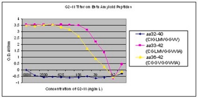
This antibody recognizes Amyloid β40 at the C-terminus.

Immunogen

Epitope: C-terminus
Linear peptide of human Amyloid β40 at the C terminus.

Application

Detect Amyloid β40 using this Anti-Amyloid β40 Antibody, clone G2-10 validated for use in WB, IH, ELISA.
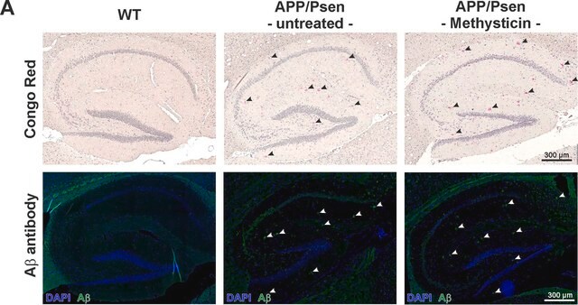
Immunohistochemistry Analysis: 1:300 dilution from a previous lot detected Amyloid β40 in Alzheimer’s diseased hippocampus tissue.
Research Category
Neuroscience
Research Sub Category
Neurodegenerative Diseases

Quality

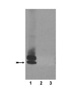
Evaluated by Western Blot in APP transgenic CRND8 mouse brain lysate.

Western Blot Analysis: 1 µg/ml of this antibody detected Amyloid β40 on 10 µg of APP transgenic CRND8 mouse brain lysate.

Target description

~ 4 kDa

Physical form

Format: Purified
Protein G Purified
Purified mouse monoclonal IgG2bκ in buffer containing 0.1 M Tris-Glycine (pH 7.4, 150 mM NaCl) with 0.05% sodium azide and 1% BSA.

Storage and Stability

Stable for 1 year at 2-8°C from date of receipt.

Analysis Note

Control
APP transgenic CRND8 mouse brain lysate

Other Notes

Concentration: Please refer to the Certificate of Analysis for the lot-specific concentration.

Disclaimer

Unless otherwise stated in our catalog or other company documentation accompanying the product(s), our products are intended for research use only and are not to be used for any other purpose, which includes but is not limited to, unauthorized commercial uses, in vitro diagnostic uses, ex vivo or in vivo therapeutic uses or any type of consumption or application to humans or animals.

Storage Class Code

12 - Non Combustible Liquids

WGK

WGK 1

Flash Point(F)

Not applicable

Flash Point(C)

Not applicable


Regulatory Listings

Regulatory Listings are mainly provided for chemical products. Only limited information can be provided here for non-chemical products. No entry means none of the components are listed. It is the user’s obligation to ensure the safe and legal use of the product.

JAN Code

MABN11:


Certificates of Analysis (COA)

Search for Certificates of Analysis (COA) by entering the products Lot/Batch Number. Lot and Batch Numbers can be found on a product’s label following the words ‘Lot’ or ‘Batch’.

Already Own This Product?

Find documentation for the products that you have recently purchased in the Document Library.

Visit the Document Library

Ge Li et al.
International journal of molecular medicine, 42(4), 1935-1944 (2018-08-08)
Aging is associated with impairment of the paravascular pathway caused by the activation of astrocytes and depolarization of protein aquaporin‑4 (AQP4) water channels, resulting in the accumulation of protein waste, including amyloid β (Aβ), in the brain parenchyma. The secreted
Ditte Z Christensen et al.
Frontiers in aging neuroscience, 6, 139-139 (2014-07-16)
Abnormalities and impairments in axonal transport are suggested to strongly contribute to the pathological alterations underlying AD. The exact mechanisms leading to axonopathy are currently unclear, but it was recently suggested that APP expression itself triggers axonal degeneration. We used
Vanessa Kurth et al.
The Journal of biological chemistry, 299(8), 104997-104997 (2023-07-03)
Presenilin-1 (PSEN1) is the catalytic subunit of the intramembrane protease γ-secretase and undergoes endoproteolysis during its maturation. Heterozygous mutations in the PSEN1 gene cause early-onset familial Alzheimer's disease (eFAD) and increase the proportion of longer aggregation-prone amyloid-β peptides (Aβ42 and/or
Jin Cui et al.
Cell discovery, 1, 15021-15021 (2015-01-01)
Despite decades of intense global effort, no disease-modifying drugs for Alzheimer's disease have emerged. Molecules targeting catalytic activities of γ-secretase or β-site APP-cleaving enzyme 1 (BACE1) have been beset by undesired side effects. We hypothesized that blocking the interaction between
Kevin Kleffman et al.
Cancer discovery, 12(5), 1314-1335 (2022-03-10)
Brain metastasis is a significant cause of morbidity and mortality in multiple cancer types and represents an unmet clinical need. The mechanisms that mediate metastatic cancer growth in the brain parenchyma are largely unknown. Melanoma, which has the highest rate

Our team of scientists has experience in all areas of research including Life Science, Material Science, Chemical Synthesis, Chromatography, Analytical and many others.

Contact Technical Service