Skip to Content
Merck
All Photos(2)

Key Documents

Safety Information

F4891

Sigma-Aldrich

Anti-Goat/Sheep IgG−FITC antibody, Mouse monoclonal

clone GT-34, purified from hybridoma cell culture

Synonym(s):

Monoclonal Anti-Goat/Sheep IgG

Sign Into View Organizational & Contract Pricing


About This Item

UNSPSC Code:
12352203
NACRES:
NA.46

biological source

mouse

Quality Level

conjugate

FITC conjugate

antibody form

purified from hybridoma cell culture

antibody product type

secondary antibodies

clone

GT-34, monoclonal

form

buffered aqueous solution

species reactivity

sheep, goat, bovine

should not react with

rat, canine, mouse, rabbit, guinea pig, chicken, horse, pig, feline, human

storage condition

protect from light

technique(s)

dot immunobinding: 1:32
immunohistochemistry (formalin-fixed, paraffin-embedded sections): 1:20
particle immunofluorescence: 1:32

isotype

IgG1

shipped in

dry ice

storage temp.

−20°C

target post-translational modification

unmodified

Looking for similar products? Visit Product Comparison Guide

Related Categories

General description

Monoclonal Anti-Goat IgG (mouse IgG1 isotype) is derived from the hybridoma produced by the fusion of mouse myeloma cells and splenocytes from an immunized mouse. IgG (immunoglobulin G) antibody subtype is a predominant serum immunoglobulins of the immune system. It is secreted by B cells and is found in blood and extracellular fluids. IgG is found in four subclasses IgG1, IgG2, IgG3 and IgG4. IgG has a monomeric structure.
The antibody reacts with an epitope located on the heavy chain of goat IgG1 and IgG2.

Specificity

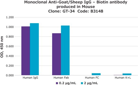
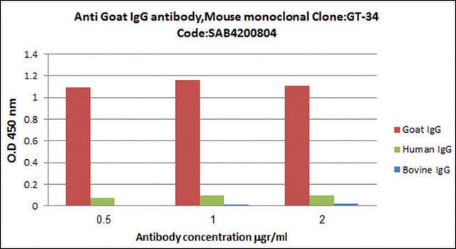
Monoclonal Anti-Goat IgG, FITC Conjugate is immunospecific for goat IgG. The antibody does not react with human, mouse, rat, rabbit, guinea pig, horse, or chicken IgGs.

Application

Biofile 39 Navya load

Biochem/physiol Actions

IgG (immunoglobulin G) antibody provides protection from infections caused by bacteria, fungi and viruses. Maternal IgG is transferred to fetus through the placenta that is vital for immune defense of the neonate against infections. IgG antibody participates in complement fixation and opsonization. Goat antibodies against many analytes are widely used in research as primary antibodies in various assay techniques. Second antibodies are that used to detect goat antibodies may lack specificity for goat immunoglobulins, and in many instances will also recognize other immunoglobulins that appear in the preparation being tested. This results in the need for extensive adsorption of the second antibody. Therefore, using a monoclonal antibody to goat IgG that is devoid of a binding capacity to other species can serve as a useful tool in many applications.

Physical form

Solution in 0.01 M sodium phosphate buffer, pH 7.4, containing 1% inactivated bovine serum albumin and 15 mM sodium azide

Storage and Stability

For continuous use, store at 2-8 °C for a maximum of one month. For extended storage, solution may be frozen in working aliquots. Repeated freezing and thawing is not recommended. Storage in "frost-free" freezers is not recommended. If slight turbidity occurs upon prolonged storage, clarify by centrifugation before use.

Disclaimer

Unless otherwise stated in our catalog or other company documentation accompanying the product(s), our products are intended for research use only and are not to be used for any other purpose, which includes but is not limited to, unauthorized commercial uses, in vitro diagnostic uses, ex vivo or in vivo therapeutic uses or any type of consumption or application to humans or animals.

Not finding the right product?  

Try our Product Selector Tool.

Storage Class Code

10 - Combustible liquids

WGK

nwg

Flash Point(F)

Not applicable

Flash Point(C)

Not applicable

Personal Protective Equipment

dust mask type N95 (US), Eyeshields, Gloves

Regulatory Listings

Regulatory Listings are mainly provided for chemical products. Only limited information can be provided here for non-chemical products. No entry means none of the components are listed. It is the user’s obligation to ensure the safe and legal use of the product.

JAN Code

F4891-VAR:
F4891-.5ML:
F4891-.2ML:
F4891-BULK:


Choose from one of the most recent versions:

Certificates of Analysis (COA)

Lot/Batch Number

Don't see the Right Version?

If you require a particular version, you can look up a specific certificate by the Lot or Batch number.

Already Own This Product?

Find documentation for the products that you have recently purchased in the Document Library.

Visit the Document Library

Bing Su et al.
The Journal of biological chemistry, 285(7), 4578-4586 (2009-12-19)
SSeCKS/Gravin/AKAP12 ("SSeCKS") encodes a cytoskeletal protein that regulates G(1) --> S progression by scaffolding cyclins, protein kinase C (PKC) and PKA. SSeCKS is down-regulated in many tumor types including prostate, and when re-expressed in MAT-LyLu (MLL) prostate cancer cells, SSeCKS
Alison McDowall et al.
The Journal of clinical investigation, 111(1), 51-60 (2003-01-04)
The adhesion receptors known as integrins perform key functions for hematopoietic cells. The platelet integrin alphaIIbbeta3 is critical in hemostasis, and the beta1 and beta2 integrins on leukocytes have many roles in cell-mediated immunity. Mutations in the beta2 subunit lead
Thomas D Barbour et al.
Kidney international, 89(4), 823-832 (2016-03-01)
C3 glomerulopathy is a complement-mediated renal disease that is frequently associated with abnormalities in regulation of the complement alternative pathway. Mice with deficiency of factor H (Cfh(-/-)), a negative alternative pathway regulator, are an established experimental model of C3 glomerulopathy
Anna L Guildford et al.
Journal of the Royal Society, Interface, 8(58), 641-649 (2010-11-26)
In-stent restenosis is a clinical complication following coronary angioplasty caused by the implantation of the metal device in the atherosclerotic vessel. Histological examination has shown a clear contribution of both inflammatory and smooth muscle cells (SMCs) to the deposition of
Alyssa C Gilmore et al.
Kidney international, 99(2), 396-404 (2020-11-02)
C3 glomerulopathy is characterized by accumulation of complement C3 within glomeruli. Causes include, but are not limited to, abnormalities in factor H, the major negative regulator of the complement alternative pathway. Factor H-deficient (Cfh-/-) mice develop C3 glomerulopathy together with

Our team of scientists has experience in all areas of research including Life Science, Material Science, Chemical Synthesis, Chromatography, Analytical and many others.

Contact Technical Service