Skip to Content
Merck
All Photos(8)

Key Documents

Safety Information

HPA010025

Sigma-Aldrich

Anti-SELENOS antibody produced in rabbit

enhanced validation

affinity isolated antibody, buffered aqueous glycerol solution

Synonym(s):

Anti-AD-015, Anti-MGC2553, Anti-SBBI8, Anti-SELS, Anti-SEPS1, Anti-VIMP

Sign Into View Organizational & Contract Pricing


About This Item

UNSPSC Code:
12352203
Human Protein Atlas Number:
NACRES:
NA.41
conjugate:
unconjugated
application:
IF
IHC
clone:
polyclonal
species reactivity:
human
citations:
17
technique(s):
immunoblotting: 0.04-0.4 μg/mL
immunofluorescence: 0.25-2 μg/mL
immunohistochemistry: 1:200-1:500

biological source

rabbit

Quality Level

conjugate

unconjugated

antibody form

affinity isolated antibody

antibody product type

primary antibodies

clone

polyclonal

form

buffered aqueous glycerol solution

species reactivity

human

enhanced validation

RNAi knockdown
Learn more about Antibody Enhanced Validation

technique(s)

immunoblotting: 0.04-0.4 μg/mL
immunofluorescence: 0.25-2 μg/mL
immunohistochemistry: 1:200-1:500

immunogen sequence

EPDVVVKRQEALAAARLKMQEELNAQVEKHKEKLKQLEEEKRRQKIEMWDSMQEGKSYKGNAKKPQEEDSPGPSTSSVLKRKSDRKPLRGGGYNPLSGEGGGACSWRPGR

shipped in

wet ice

storage temp.

−20°C

target post-translational modification

unmodified

Gene Information

human ... SELS(55829)

General description

Selenoprotein S (SELS) is a transmembrane protein that is composed of 189 amino acids. Alternative splicing produces two isoforms of this enzyme, which are different at their 3′UTR (untranslated region) sequences. Both these isoforms are widely expressed. It is one of the most widely present selenoproteins in eukaryotes. This protein is localized to the ER (endoplasmic reticulum).

Immunogen

selenoprotein S recombinant protein epitope signature tag (PrEST)

Application

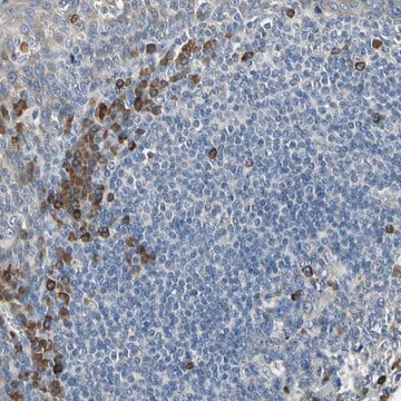
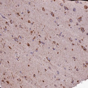
Anti-SELS antibody produced in rabbit, a Prestige Antibody, is developed and validated by the Human Protein Atlas (HPA) project . Each antibody is tested by immunohistochemistry against hundreds of normal and disease tissues. These images can be viewed on the Human Protein Atlas (HPA) site by clicking on the Image Gallery link. The antibodies are also tested using immunofluorescence and western blotting. To view these protocols and other useful information about Prestige Antibodies and the HPA, visit sigma.com/prestige.
Applications in which this antibody has been used successfully, and the associated peer-reviewed papers, are given below.
Immunofluorescence (1 paper)
Western Blotting (1 paper)

Biochem/physiol Actions

Selenoprotein S (SELS) plays a crucial, though unidentified role in unfolded protein response. It is thought to act as a reductase. The levels of this protein are regulated by circulating glucose and insulin levels, which share an inverse relation with each other. Polymorphism -105G>A in this gene is linked with increased susceptibility to Kashin-Beck disease (KBD), where it regulates the expression of expression of PI3K (phosphatidylinositol 3-kinase)/Akt signaling pathway. It plays an essential role in the production of inflammatory cytokines. In mice, it might confer protection against LPS (lipopolysaccharide)-induced sepsis and organ damage. Therefore, it might have potential as a therapeutic target LPS-induced sepsis. Variants present in the promoter region of SELS are linked with increased susceptibility to Hashimoto′s thyroiditis.

Features and Benefits

Prestige Antibodies® are highly characterized and extensively validated antibodies with the added benefit of all available characterization data for each target being accessible via the Human Protein Atlas portal linked just below the product name at the top of this page. The uniqueness and low cross-reactivity of the Prestige Antibodies® to other proteins are due to a thorough selection of antigen regions, affinity purification, and stringent selection. Prestige antigen controls are available for every corresponding Prestige Antibody and can be found in the linkage section.

Every Prestige Antibody is tested in the following ways:
  • IHC tissue array of 44 normal human tissues and 20 of the most common cancer type tissues.
  • Protein array of 364 human recombinant protein fragments.

Linkage

Corresponding Antigen APREST71935

Physical form

Solution in phosphate-buffered saline, pH 7.2, containing 40% glycerol and 0.02% sodium azide

Legal Information

Prestige Antibodies is a registered trademark of Merck KGaA, Darmstadt, Germany

Disclaimer

Unless otherwise stated in our catalog or other company documentation accompanying the product(s), our products are intended for research use only and are not to be used for any other purpose, which includes but is not limited to, unauthorized commercial uses, in vitro diagnostic uses, ex vivo or in vivo therapeutic uses or any type of consumption or application to humans or animals.

Not finding the right product?  

Try our Product Selector Tool.

Storage Class Code

10 - Combustible liquids

WGK

WGK 1

Personal Protective Equipment

dust mask type N95 (US), Eyeshields, Gloves

Regulatory Listings

Regulatory Listings are mainly provided for chemical products. Only limited information can be provided here for non-chemical products. No entry means none of the components are listed. It is the user’s obligation to ensure the safe and legal use of the product.

JAN Code

HPA010025-100UL:
HPA010025-25UL:


Choose from one of the most recent versions:

Certificates of Analysis (COA)

Lot/Batch Number

Don't see the Right Version?

If you require a particular version, you can look up a specific certificate by the Lot or Batch number.

Already Own This Product?

Find documentation for the products that you have recently purchased in the Document Library.

Visit the Document Library

Craig Robert Wright et al.
Mediators of inflammation, 2017, 7043429-7043429 (2017-06-09)
Excessive inflammation is a hallmark of muscle myopathies, including Duchenne muscular dystrophy (DMD). There is interest in characterising novel genes that regulate inflammation due to their potential to modify disease progression. Gene polymorphisms in Selenoprotein S (Seps1) are associated with
X A Du et al.
Osteoarthritis and cartilage, 23(2), 210-216 (2014-11-30)
To investigate the relationship between SEPS1 polymorphism and phosphatidylinositol 3-kinase (PI3K)/Akt signaling pathway in Kashin-Beck disease (KBD) and further explore the pathogenesis of KBD. Polymerase chain reaction-restriction fragment length polymorphism (PCR-RFLP) was used to detect SEPS1 -105G>A polymorphism in 232
Lei He et al.
Molecular medicine reports, 9(5), 1869-1876 (2014-02-28)
Septic shock and sequential multiple organ failure are the main cause of mortality in patients with sepsis. The induction of inflammation during sepsis is a complex biological cascade, which requires successful therapeutic intervention. Selenoprotein S (SEPS1) is a novel endoplasmic
Alex B Addinsall et al.
American journal of physiology. Regulatory, integrative and comparative physiology, 315(2), R380-R396 (2018-04-19)
Selenoprotein S (Seps1) is an endoplasmic reticulum (ER) resident antioxidant implicated in ER stress and inflammation. In human vastus lateralis and mouse hindlimb muscles, Seps1 localization and expression were fiber-type specific. In male Seps1+/- heterozygous mice, spontaneous physical activity was
Liliana R Santos et al.
The Journal of clinical endocrinology and metabolism, 99(4), E719-E723 (2014-01-30)
The association between selenium and inflammation and the relevance of selenoproteins in follicular thyroid cell physiology have pointed to a putative role of selenoproteins in the pathogenesis of autoimmune thyroid diseases. The aim of this study was to evaluate the

Our team of scientists has experience in all areas of research including Life Science, Material Science, Chemical Synthesis, Chromatography, Analytical and many others.

Contact Technical Service