Skip to Content
Merck
All Photos(1)

Key Documents

Safety Information

SAB4200671

Sigma-Aldrich

Anti-S-100 (β-Subunit) antibody, Mouse monoclonal

clone SH-B1, purified from hybridoma cell culture

Synonym(s):

Monoclonal Anti-S-100 (β-Subunit) antibody produced in mouse, NEF, S100, S100-B, S100beta

Sign Into View Organizational & Contract Pricing


About This Item

UNSPSC Code:
12352203
NACRES:
NA.46

biological source

mouse

Quality Level

antibody form

purified from hybridoma cell culture

antibody product type

primary antibodies

clone

SH-B1, monoclonal

species reactivity

cat, rabbit, porcine, bovine, rat, human

concentration

~1 mg/mL

technique(s)

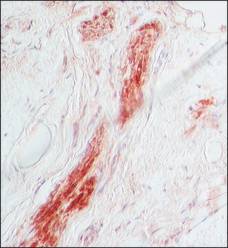
immunohistochemistry: 1.5-3  μg/mL using immunoperoxidase labeling of pronase digested, formalin-fixed, paraffin-embedded sections of rabbit tongue.

isotype

IgG1

shipped in

dry ice

storage temp.

−20°C

target post-translational modification

unmodified

Gene Information

human ... S100B(6285)

General description

Monoclonal Anti-S-100 (β-subunit) (mouse IgG1 isotype) is derived from the SH-B1 hybridoma produced by the fusion of mouse myeloma cells and splenocytes from an immunized mouse. S-100 is a set of small, thermolabile, highly acidic homo or hetero-dimer calcium binding proteins. The protein exists in two isoforms namely, S-100α and S-100β, which are brain specific.
S-100β is a calcium binding protein. It is mainly present in astrocytes and neurons of hindbrain and spinal cord.

Immunogen

Purified bovine brain S-100β

Application

Monoclonal Anti-S-100 (β-Subunit) antibody produced in mouse has been used in:
  • immunohistochemistry
  • enzyme linked immunosorbent assay (ELISA) (Ca2+ ion independent)
  • immunocytochemistry
  • immunoblotting
  • dot blot
  • immunohistochemistry.

Biochem/physiol Actions

S-100 is involved in cell-growth regulation, increasing membrane permeability to cations, inflammatory response in many brain diseases, including schizophrenia, stimulation of nucleolar RNA polymerase activity and transporting proteins and free fatty acids in adipocytes. S-100β tissue distribution can be a useful tool in the differential diagnosis of neoplasms and proliferative processes.
S-100β protein interacts with synaptic, cytoskeletal and cell cycle proteins. Additionally, it can regulate calcium levels in glial and neuronal cells. It is involved in neuronal plasticity, astrogliosis and neuronal cell survival. S-100β is associated with Alzheimer disease and amyotrophic lateral sclerosis.

Disclaimer

Unless otherwise stated in our catalog or other company documentation accompanying the product(s), our products are intended for research use only and are not to be used for any other purpose, which includes but is not limited to, unauthorized commercial uses, in vitro diagnostic uses, ex vivo or in vivo therapeutic uses or any type of consumption or application to humans or animals.

Not finding the right product?  

Try our Product Selector Tool.

Storage Class Code

10 - Combustible liquids

Flash Point(F)

Not applicable

Flash Point(C)

Not applicable


Regulatory Listings

Regulatory Listings are mainly provided for chemical products. Only limited information can be provided here for non-chemical products. No entry means none of the components are listed. It is the user’s obligation to ensure the safe and legal use of the product.

JAN Code

SAB4200671-100UL:
SAB4200671-BULK:
SAB4200671-VAR:


Certificates of Analysis (COA)

Search for Certificates of Analysis (COA) by entering the products Lot/Batch Number. Lot and Batch Numbers can be found on a product’s label following the words ‘Lot’ or ‘Batch’.

Already Own This Product?

Find documentation for the products that you have recently purchased in the Document Library.

Visit the Document Library

A Migheli et al.
Neuroscience letters, 261(1-2), 25-28 (1999-03-19)
Amyotrophic lateral sclerosis (ALS) is a neurodegenerative disease characterized by progressive motor neuron loss and astrogliosis. We studied the immunohistochemical expression of S-100beta, a calcium-binding protein with both neurotrophic and neurotoxic activities, in the spinal cord of patients with ALS.
Postnatal activation of TLR4 in astrocytes promotes excitatory synaptogenesis in hippocampal neurons
Shen Y, et al.
The Journal of cell biology, 215(5), 719-734 (2016)
Yan Guo et al.
Signal transduction and targeted therapy, 9(1), 32-32 (2024-02-14)
The appropriate and specific response of nerve cells to various external cues is essential for the establishment and maintenance of neural circuits, and this process requires the proper recruitment of adaptor molecules to selectively activate downstream pathways. Here, we identified
Marc Oria et al.
Frontiers in molecular neuroscience, 15, 888351-888351 (2022-07-06)
During embryonic spinal cord development, neural progenitor cells (NPCs) generate three major cell lines: neurons, oligodendrocytes, and astrocytes at precise times and locations within the spinal cord. Recent studies demonstrate early astrogenesis in animal models of spina bifida, which may
Clinicoradiological characteristics, management and prognosis of primary myeloid sarcoma of the central nervous system: A report of four cases.
Yang B, et al.
Oncology Letters, 14(3), 3825-3831 (2017)

Our team of scientists has experience in all areas of research including Life Science, Material Science, Chemical Synthesis, Chromatography, Analytical and many others.

Contact Technical Service