Skip to Content
Merck
All Photos(2)

Key Documents

Safety Information

SAB4301138

Sigma-Aldrich

Anti-GFP antibody produced in rabbit

affinity isolated antibody

Synonym(s):

GFP-like chromoprotein

Sign Into View Organizational & Contract Pricing


About This Item

UNSPSC Code:
12352203
NACRES:
NA.41
clone:
polyclonal
application:
WB
technique(s):
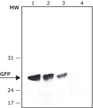
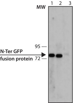
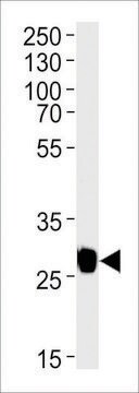
western blot: 1:1000-1:10000 (Cell Lysate)
citations:
61

biological source

rabbit

Quality Level

antibody form

affinity isolated antibody

antibody product type

primary antibodies

clone

polyclonal

form

buffered aqueous solution

mol wt

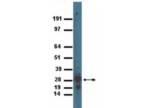
27 kDa

concentration

3.2 mg/mL

technique(s)

western blot: 1:1000-1:10000 (Cell Lysate)

isotype

IgG

shipped in

wet ice

storage temp.

−20°C

target post-translational modification

unmodified

General description

Green fluorescent protein (GFP) is a 27kDa protein, derived from the bioluminescent jellyfish Aequorea victoria, in which light is produced when energy is transferred from the Ca2+- activated photoprotein aequorin to GFP.

Specificity

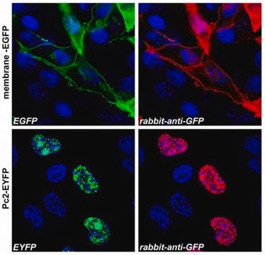
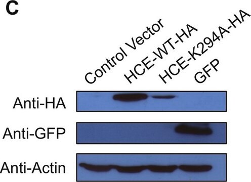
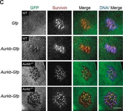
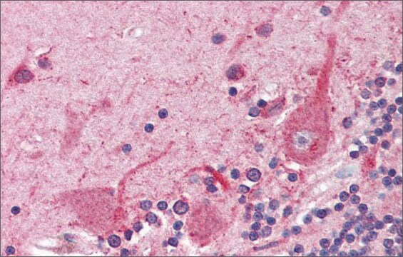
The antibody detects transfected proteins containing GFP tag.

Immunogen

Full length fusion protein

Application

Anti-GFP antibody produced in rabbit has been used in:
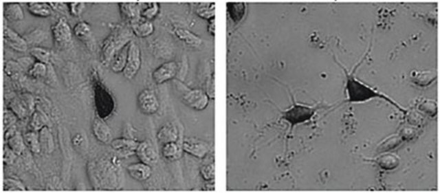
  • double immunofluorescence staining
  • immunoblot analysis.
  • immunoprecipitation.

Biochem/physiol Actions

Green fluorescent protein (GFP) is a reporter molecule which is used for checking gene expression and protein localization in vivo, in situ and in real time. GFP emits green light when it is excited with UV/blue light. The GFP fluorescence remains stable and can be detected non-invasively in living cells. GFP is considered as a unique tool to monitor dynamic processes in several living cells or organisms. When expressed in either eukaryotic or prokaryotic cells and illuminated by blue or UV light, GFP yields a bright green fluorescence.

Features and Benefits

Evaluate our antibodies with complete peace of mind. If the antibody does not perform in your application, we will issue a full credit or replacement antibody. Learn more.

Physical form

Rabbit IgG in pH7.3 PBS, 0.05% NaN3, 50% Glycerol.

Disclaimer

Unless otherwise stated in our catalog or other company documentation accompanying the product(s), our products are intended for research use only and are not to be used for any other purpose, which includes but is not limited to, unauthorized commercial uses, in vitro diagnostic uses, ex vivo or in vivo therapeutic uses or any type of consumption or application to humans or animals.

Not finding the right product?  

Try our Product Selector Tool.

Storage Class Code

10 - Combustible liquids

WGK

WGK 1

Flash Point(F)

Not applicable

Flash Point(C)

Not applicable


Regulatory Listings

Regulatory Listings are mainly provided for chemical products. Only limited information can be provided here for non-chemical products. No entry means none of the components are listed. It is the user’s obligation to ensure the safe and legal use of the product.

JAN Code

SAB4301138-100UL:


Choose from one of the most recent versions:

Certificates of Analysis (COA)

Lot/Batch Number

Don't see the Right Version?

If you require a particular version, you can look up a specific certificate by the Lot or Batch number.

Already Own This Product?

Find documentation for the products that you have recently purchased in the Document Library.

Visit the Document Library

UGT79B31 is responsible for the final modification step of pollen-specific flavonoid biosynthesis in Petunia hybrida.
Knoch E, et al.
Planta, 247(4), 779-790 (2018)
Minna M Koskela et al.
The Plant cell, 30(8), 1695-1709 (2018-07-04)
The amount of light energy received by the photosynthetic reaction centers photosystem II (PSII) and photosystem I (PSI) is balanced through state transitions. Reversible phosphorylation of a light-harvesting antenna trimer (L-LHCII) orchestrates the association between L-LHCII and the photosystems, thus
RNA-dependent RNA polymerases of both virulent and benign rabbit caliciviruses induce striking rearrangement of Golgi membranes.
Urakova N, et al.
PLoS ONE, 12(1), e0169913-e0169913 (2017)
Green fluorescent protein as a reporter for macromolecular localization in bacterial cells.
Margolin W
Methods, 20(1), 62-72 (2000)
David A Rhodes et al.
Frontiers in immunology, 9, 662-662 (2018-04-20)
Activation of human Vγ9/Vδ2 T cells by "phosphoantigens" (pAg), the microbial metabolite (E)-4-hydroxy-3-methyl-but-2-enyl pyrophosphate (HMB-PP) and the endogenous isoprenoid intermediate isopentenyl pyrophosphate, requires expression of butyrophilin BTN3A molecules by presenting cells. However, the precise mechanism of activation of Vγ9/Vδ2 T

Our team of scientists has experience in all areas of research including Life Science, Material Science, Chemical Synthesis, Chromatography, Analytical and many others.

Contact Technical Service