Skip to Content
Merck
All Photos(3)

Key Documents

SAB4501665

Sigma-Aldrich

Anti-Keratin 18 antibody produced in rabbit

affinity isolated antibody

Synonym(s):

CK 18, CK18, CYK18, Cytokeratin endo B, K18

Sign Into View Organizational & Contract Pricing


About This Item

UNSPSC Code:
12352203
NACRES:
NA.41
conjugate:
unconjugated
application:
ELISA
IF
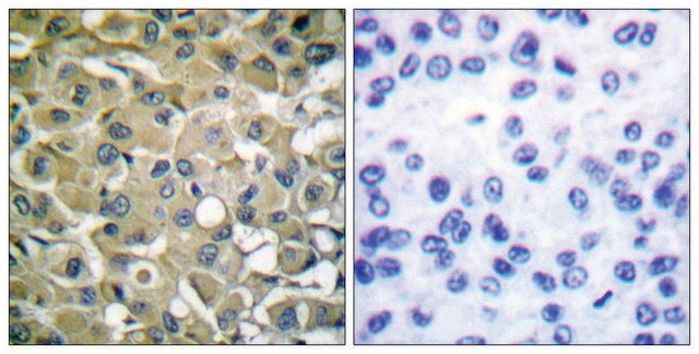
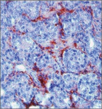
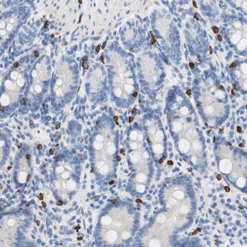
IHC
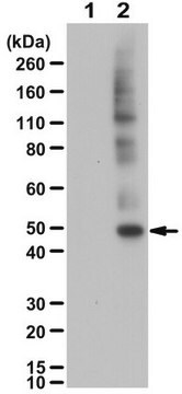
WB
clone:
polyclonal
species reactivity:
mouse, rat, human
citations:
7
technique(s):
ELISA: 1:10000
immunofluorescence: 1:100-1:500
immunohistochemistry: 1:50-1:100
western blot: 1:500-1:1000

biological source

rabbit

Quality Level

conjugate

unconjugated

antibody form

affinity isolated antibody

antibody product type

primary antibodies

clone

polyclonal

form

buffered aqueous solution

mol wt

antigen 48 kDa

species reactivity

mouse, rat, human

concentration

~1 mg/mL

technique(s)

ELISA: 1:10000
immunofluorescence: 1:100-1:500
immunohistochemistry: 1:50-1:100
western blot: 1:500-1:1000

NCBI accession no.

UniProt accession no.

shipped in

wet ice

storage temp.

−20°C

target post-translational modification

unmodified

Gene Information

human ... KRT18(3875)

General description

Anti-Keratin 18 antibody detects endogenous levels of total Keratin 18 protein.
Keratin 18/cytokeratin 18 (CK18) is a structural protein. It is present in single-layer epithelia. It is located on human chromosome 12q13.

Immunogen

The antiserum was produced against synthesized peptide derived from human Keratin 18.

Immunogen Range: 381-430

Biochem/physiol Actions

Keratins help to maintain the structural integrity. They also control Fas‐mediated apoptosis and regulate cell size and protein synthesis. Keratin 18 plays a major role in the development of lung cancer. It acts as a therapeutic target for non-small cell lung cancer (NSCLC).

Features and Benefits

Evaluate our antibodies with complete peace of mind. If the antibody does not perform in your application, we will issue a full credit or replacement antibody. Learn more.

Physical form

Rabbit IgG in phosphate buffered saline (without Mg2+ and Ca2+), pH 7.4, 150mM NaCl, 0.02% sodium azide and 50% glycerol.

Disclaimer

Unless otherwise stated in our catalog or other company documentation accompanying the product(s), our products are intended for research use only and are not to be used for any other purpose, which includes but is not limited to, unauthorized commercial uses, in vitro diagnostic uses, ex vivo or in vivo therapeutic uses or any type of consumption or application to humans or animals.

Not finding the right product?  

Try our Product Selector Tool.

Storage Class Code

10 - Combustible liquids

WGK

nwg

Flash Point(F)

Not applicable

Flash Point(C)

Not applicable


Choose from one of the most recent versions:

Certificates of Analysis (COA)

Lot/Batch Number

Don't see the Right Version?

If you require a particular version, you can look up a specific certificate by the Lot or Batch number.

Already Own This Product?

Find documentation for the products that you have recently purchased in the Document Library.

Visit the Document Library

Lucy Pilcher et al.
Stem cells international, 2024, 8601360-8601360 (2024-01-19)
The epicardium is a layer of mesothelial cells that covers the surface of the heart. During development, epicardial cells undergo epithelial-to-mesenchymal transition (EMT) to form multipotent precursors that migrate into the heart and contribute to the coronary vasculature by differentiating
Diptendu Chatterjee et al.
European heart journal, 41(30), 2878-2890 (2020-06-14)
Brugada syndrome (BrS) is characterized by a unique electrocardiogram (ECG) pattern and life-threatening arrhythmias. However, the Type 1 Brugada ECG pattern is often transient, and a genetic cause is only identified in <25% of patients. We sought to identify an
Kasey Y C Lau et al.
Cell stem cell, 29(10), 1445-1458 (2022-09-10)
Several in vitro models have been developed to recapitulate mouse embryogenesis solely from embryonic stem cells (ESCs). Despite mimicking many aspects of early development, they fail to capture the interactions between embryonic and extraembryonic tissues. To overcome this difficulty, we have
Carlos A Pinzón-Arteaga et al.
Cell stem cell, 30(5), 611-616 (2023-05-06)
Understanding the mechanisms of blastocyst formation and implantation is critical for improving farm animal reproduction but is hampered by a limited supply of embryos. Here, we developed an efficient method to generate bovine blastocyst-like structures (termed blastoids) via assembling bovine
Characterization of new members of the human type II keratin gene family and a general evaluation of the keratin gene domain on chromosome 12q13.13
Rogers MA, et al.
The Journal of Investigative Dermatology, 124(3), 536-544 (2005)

Our team of scientists has experience in all areas of research including Life Science, Material Science, Chemical Synthesis, Chromatography, Analytical and many others.

Contact Technical Service