Skip to Content
Merck
All Photos(3)

Key Documents

SAB4502149

Sigma-Aldrich

Anti-PDGF Receptor β antibody produced in rabbit

affinity isolated antibody

Synonym(s):

CD140b, PDGF-R-β, PDGFR, PDGFR-β, kinase PDGFR-β

Sign Into View Organizational & Contract Pricing


About This Item

MDL number:
UNSPSC Code:
12352203
NACRES:
NA.41

biological source

rabbit

Quality Level

conjugate

unconjugated

antibody form

affinity isolated antibody

antibody product type

primary antibodies

clone

polyclonal

form

buffered aqueous solution

mol wt

antigen 123 kDa

species reactivity

rat, mouse, human

concentration

~1 mg/mL

technique(s)

ELISA: 1:40000
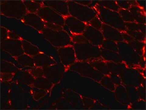
immunofluorescence: 1:100-1:500
immunohistochemistry: 1:50-1:100
western blot: 1:500-1:1000

NCBI accession no.

UniProt accession no.

shipped in

wet ice

storage temp.

−20°C

target post-translational modification

unmodified

Gene Information

human ... PDGFRB(5159)

General description

The PDGFRB (platelet-derived growth factor receptor β) gene is mapped to human chromosome 5q32. The encoded protein is a cell surface tyrosine kinase receptor. Anti-PDGF receptor β antibody detects endogenous levels of total PDGF receptor β protein.

Immunogen

The antiserum was produced against synthesized peptide derived from human PDGF Receptor beta.

Immunogen Range: 718-767

Biochem/physiol Actions

PDGFRB (platelet-derived growth factor β) has its key role in cell proliferation, differentiation and survival. It also mediates cell movement and tissue repair mechanism. PDGFβ functions as a mitogen, by binding to the PDGF receptor β. Variation in PDGFRB gene leads to a number of cancer types including glioblastoma, dermatofibrosarcoma, medulloblastoma and endocrine pancreatic tumor. PDGFR-β signaling pathway, can thus serve as an important target for cancer therapy.

Features and Benefits

Evaluate our antibodies with complete peace of mind. If the antibody does not perform in your application, we will issue a full credit or replacement antibody. Learn more.

Physical form

Rabbit IgG in phosphate buffered saline (without Mg2+ and Ca2+), pH 7.4, 150mM NaCl, 0.02% sodium azide and 50% glycerol.

Disclaimer

Unless otherwise stated in our catalog or other company documentation accompanying the product(s), our products are intended for research use only and are not to be used for any other purpose, which includes but is not limited to, unauthorized commercial uses, in vitro diagnostic uses, ex vivo or in vivo therapeutic uses or any type of consumption or application to humans or animals.

Not finding the right product?  

Try our Product Selector Tool.

Storage Class Code

10 - Combustible liquids

WGK

nwg

Flash Point(F)

Not applicable

Flash Point(C)

Not applicable


Choose from one of the most recent versions:

Certificates of Analysis (COA)

Lot/Batch Number

Don't see the Right Version?

If you require a particular version, you can look up a specific certificate by the Lot or Batch number.

Already Own This Product?

Find documentation for the products that you have recently purchased in the Document Library.

Visit the Document Library

The 3?-end region of the human PDGFR-beta core promoter nuclease hypersensitive element forms a mixture of two unique end-insertion G-quadruplexes
Onel B, et al.
Biochim. Biophys. Acta Gen. Subj., 1862(4), 846-854 (2018)
Tyrosines-740/751 of PDGFRbeta contribute to the activation of Akt/Hif1alphaTGFbeta nexus to drive high glucose-induced glomerular mesangial cell hypertrophy
Das F, et al.
Cellular Signalling, 42(6), 44-53 (2018)
Mutations in PDGFRB cause autosomal-dominant infantile myofibromatosis.
Martignetti J A, et al.
American Journal of Human Genetics, 92(6), 1001-1007 (2013)
Nigella sativa L. seed extracts promote wound healing progress by activating VEGF and PDGF signaling pathways: An in vitro and in silico study.
Palanisamy, et al.
F1000Research, 12, 436-436 (2023)

Our team of scientists has experience in all areas of research including Life Science, Material Science, Chemical Synthesis, Chromatography, Analytical and many others.

Contact Technical Service