32922
Lugol solution
according to Lugol
Synonym(s):
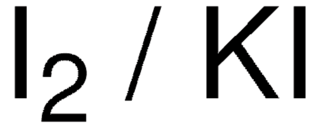
Iodine/Potassium iodide solution
Sign Into View Organizational & Contract Pricing
All Photos(1)
About This Item
Recommended Products
form
liquid
Quality Level
quality
according to Lugol
density
1.006 g/cm3 at 20 °C
application(s)
diagnostic assay manufacturing
food and beverages
hematology
histology
storage temp.
room temp
SMILES string
[K+].[I-].II
InChI
1S/I2.HI.K/c1-2;;/h;1H;/q;;+1/p-1
InChI key
XUEKKCJEQVGHQZ-UHFFFAOYSA-M
Looking for similar products? Visit Product Comparison Guide
Related Categories
General description
Lugol′s solution is a ready-to-use solution that comprises 6% iodine in 4% potassium iodide. It is commonly used as a mordant for Gram staining in bacteriology. It is used for staining bacterial target structures for diagnostic purposes by fixing, staining, counterstaining, and mounting bacteriological specimen materials.
Application
- Lugol′s solution is generally used as a mordant in Gram staining, and for fixing invertebrate blood cells and protozoa.
- It is suitable for the detection of starch in plants.
- It has been used in a study to assess narrow-band imaging without image magnification for detecting high-grade dysplasia and intramucosal esophageal squamous cell carcinoma.
- It has been used in a study to determine its benefit in aiding the detection, presence, and spread of small squamous cell carcinomas of the esophagus.
Biochem/physiol Actions
Iodine diffuses rapidly through hydrophilic and hydrophobic domains of tissues and loosely binds to proteins, staining them brown. It reacts irreversibly with unsaturated lipid linkages and with tyrosine and histidine aromatic rings.
Features and Benefits
- Ready-to-use solution.
Principle
In bacteriology, Gram staining allows rapid differentiation between Gram-positive and Gram-negative bacteria. The mureine structure of the bacterial cell wall is the basis of the color affinity. Bacteria are first stained with the primary stain, crystal violet. Lugol solution is added thereafter as the mordant, forming crystal violet-iodine complexes. During the decolorization, these complexes stay entrapped in the multilayer mureine structure of the cell wall of Gram-positive bacteria which appear blue-violet. Gram-negative bacteria have a single-layered cell wall that does not retain the dye-iodine complexes and are counterstained with safranine solution, appearing pink.
Storage Class Code
10 - Combustible liquids
WGK
WGK 2
Flash Point(F)
Not applicable
Flash Point(C)
Not applicable
Personal Protective Equipment
dust mask type N95 (US), Eyeshields, Gloves
Choose from one of the most recent versions:
Already Own This Product?
Find documentation for the products that you have recently purchased in the Document Library.
Histological and Histochemical Methods: Theory and Practice
J.A. Kiernan
Histological and Histochemical Methods: Theory and Practice (2015)
Jason M Cole et al.
Proceedings of the National Academy of Sciences of the United States of America, 115(25), 6335-6340 (2018-06-07)
In the field of X-ray microcomputed tomography (μCT) there is a growing need to reduce acquisition times at high spatial resolution (approximate micrometers) to facilitate in vivo and high-throughput operations. The state of the art represented by synchrotron light sources
Our team of scientists has experience in all areas of research including Life Science, Material Science, Chemical Synthesis, Chromatography, Analytical and many others.
Contact Technical Service


