F3777
Anti-Actin, α-Smooth Muscle - FITC antibody, Mouse monoclonal
clone 1A4, purified from hybridoma cell culture
Synonym(s):
Monoclonal Anti-Actin, α-Smooth Muscle, alpha SMA-FITC antibody, alpha smooth muscle actin - FITC antibody, SMA
About This Item
Recommended Products
biological source
mouse
Quality Level
conjugate
FITC conjugate
antibody form
purified from hybridoma cell culture
antibody product type
primary antibodies
clone
1A4, monoclonal
form
buffered aqueous solution
mol wt
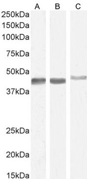
antigen ~42 kDa
species reactivity
human, mouse, rat, chicken, frog, canine, rabbit, guinea pig, goat, bovine, sheep, snake
technique(s)
immunohistochemistry (formalin-fixed, paraffin-embedded sections): 1:500 using human tonsil or appendix
isotype
IgG2a
UniProt accession no.
shipped in
dry ice
storage temp.
−20°C
target post-translational modification
unmodified
Gene Information
human ... ACTA2(59)
mouse ... Acta2(11475)
rat ... Acta2(81633)
Looking for similar products? Visit Product Comparison Guide
General description
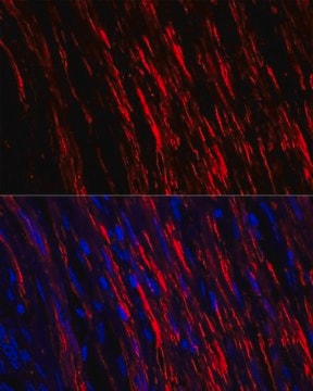
The antibody reacts with normal and neoplastic, human vascular and visceral smooth muscle cells. It reacts with normal myoepithelial cells, pericytes, eye lens cells, hair follicle cells and certain stromal cells in the intestine, testis, lymphoid organs, liver, ovary and bone marrow. The antibody also reacts with stromal myofibroblasts in hypertrophic scars, and in neoplastic tissues. α-smooth muscle actin is transiently co-expressed with sarcomeric α-actin during myogenesis in chicken and rat embryos. It was also found in the ventricular conducting tract of adult mammalian heart.
Specificity
Immunogen
Application
- immunohistochemical (IHC) staining of heart, lung, kidney, and liver of mice to examine the PHD2 deficiency in internal organs
- immunostaining to analyze vascular smooth muscle cells/pericytes development in platelet-derived growth factor (PDGFR) and PDGFR- β-/- embryos
- immunofluorescent analysis of implanted cells, frozen tissue samples to study the similar levels of expression of SMaA and SM-MHC as established primary SMC lines
- western blots of lens epithelial cells to determine the distribution of α-SMA expression in lens epithelial cell
- Immunohistochemical examination of lesion morphology
- flow cytometry
- immunohistochemistry, proliferation and TUNEL (terminal deoxynucleotidyl transferase (TdT) dUTP nick-end labeling) assays
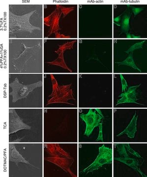
- immunocytochemistry
IHC analysis of x-gal stained muouse cardiac tissue was performed using the primary antibody, mouse monoclonal anti-smooth muscle actin to identify myofibroblasts.
Biochem/physiol Actions
Physical form
Storage and Stability
Other Notes
Disclaimer
Not finding the right product?
Try our Product Selector Tool.
Storage Class Code
12 - Non Combustible Liquids
WGK
nwg
Flash Point(F)
Not applicable
Flash Point(C)
Not applicable
Personal Protective Equipment
Choose from one of the most recent versions:
Certificates of Analysis (COA)
Don't see the Right Version?
If you require a particular version, you can look up a specific certificate by the Lot or Batch number.
Already Own This Product?
Find documentation for the products that you have recently purchased in the Document Library.
Customers Also Viewed
Our team of scientists has experience in all areas of research including Life Science, Material Science, Chemical Synthesis, Chromatography, Analytical and many others.
Contact Technical Service


