GBL654008
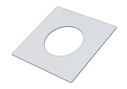
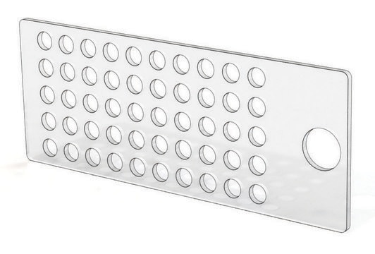
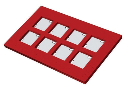
Grace Bio-Labs SecureSeal™ imaging spacer
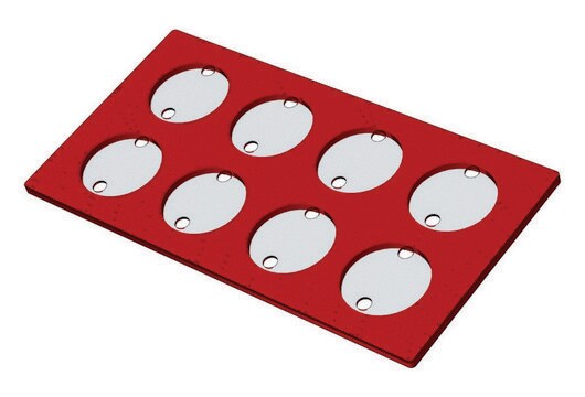
8 wells, diam. × thickness 9 mm × 0.12 mm
Synonym(s):
peal and stick adhesive chamber spacers, thin adhesive spacers for coverslpis, thin adhesive spacers for slides
Sign Into View Organizational & Contract Pricing
All Photos(2)
About This Item
UNSPSC Code:
41122600
NACRES:
NB.22
Recommended Products
material
adhesive/adhesive
colorless
sterility
non-sterile
packaging
pack of 100 ea
manufacturer/tradename
Grace Bio-Labs 654008
diam. × thickness
9 mm × 0.12 mm
external L × W
25 mm × 25 mm
wells
8
Looking for similar products? Visit Product Comparison Guide
General description
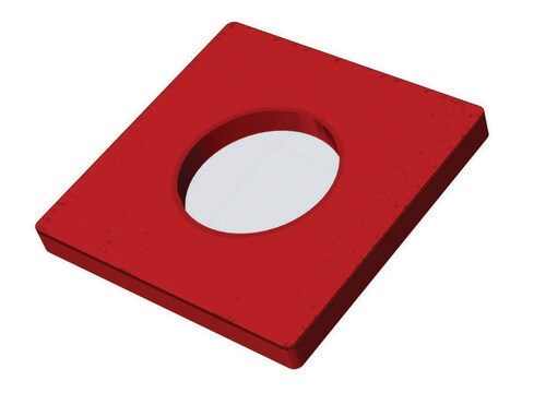
Imaging spacers are ultra-thin adhesive spacers which peel-and-stick to coverglass or microscope slides to confine specimens without compression. Layer multiple spacers to custom build chambers to any depth desired.
Application
Imaging
Microscopy
High-temperature single-molecule kinetic analysis
anti-Stokes Raman scattering microscopy
Microscopy
High-temperature single-molecule kinetic analysis
anti-Stokes Raman scattering microscopy
Legal Information
SecureSeal is a trademark of Grace Bio-Labs, Inc.
Choose from one of the most recent versions:
Certificates of Analysis (COA)
Lot/Batch Number
Sorry, we don't have COAs for this product available online at this time.
If you need assistance, please contact Customer Support.
Already Own This Product?
Find documentation for the products that you have recently purchased in the Document Library.
Customers Also Viewed
Zhixing Chen et al.
Journal of the American Chemical Society, 136(22), 8027-8033 (2014-05-23)
Vibrational imaging such as Raman microscopy is a powerful technique for visualizing a variety of molecules in live cells and tissues with chemical contrast. Going beyond the conventional label-free modality, recent advance of coupling alkyne vibrational tags with stimulated Raman
Karl-Frédéric Vieux et al.
Scientific reports, 8(1), 6812-6812 (2018-05-03)
In many cell types, the length of the poly(A) tail of an mRNA is closely linked to its fate - a long tail is associated with active translation, a short tail with silencing and degradation. During mammalian oocyte development, two
Laleh Abbassi et al.
Nature communications, 12(1), 1438-1438 (2021-03-06)
Germ cells are physically coupled to somatic support cells of the gonad during differentiation, but this coupling must be disrupted when they are mature, freeing them to participate in fertilization. In mammalian females, coupling occurs via specialized filopodia that project
Stephany El-Hayek et al.
Biology of reproduction, 93(2), 47-47 (2015-06-13)
Germ cells develop in intimate contact and communication with somatic cells of the gonad. In female mammals, oocyte development depends crucially on gap junctions that couple it to the surrounding somatic granulosa cells of the follicle, yet the mechanisms that
Our team of scientists has experience in all areas of research including Life Science, Material Science, Chemical Synthesis, Chromatography, Analytical and many others.
Contact Technical Service