Skip to Content
Merck
All Photos(1)

Key Documents

S0664

Sigma-Aldrich

Monoclonal Anti-Syntaxin antibody produced in mouse

clone HPC-1, ascites fluid

Synonym(s):

Anti-HPC-1, Anti-P35-1, Anti-STX1, Anti-SYN1A

Sign Into View Organizational & Contract Pricing


About This Item

MDL number:
UNSPSC Code:
12352203
NACRES:
NA.41

biological source

mouse

Quality Level

conjugate

unconjugated

antibody form

ascites fluid

antibody product type

primary antibodies

clone

HPC-1, monoclonal

mol wt

antigen 35 kDa

contains

15 mM sodium azide

species reactivity

rat, bovine, rabbit

technique(s)

immunocytochemistry: suitable using monolayer cultures of neonatal retina cells
microarray: suitable
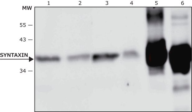
western blot: 1:2,000 using crude preparation of synaptic vesicles from rat cerebral cortex

isotype

IgG1

UniProt accession no.

shipped in

dry ice

storage temp.

−20°C

target post-translational modification

unmodified

Gene Information

Related Categories

General description

Monoclonal Anti-Syntaxin (mouse IgG1 isotype) is derived from the hybridoma produced by the fusion of mouse myeloma cells and splenocytes from an immunized mouse. Syntaxin (also cited as HPC-1 antigen), a 35 kDa molecule with carboxyl-terminal membrane anchor is a synaptic protein. Syntaxin 1 is a membrane protein. Neurons express syntaxin 1A.
Syntaxin is a 35kD synaptic protein capable of interacting with the synaptic vesicle protein synaptotagmin. It facilitates the docking and fusion of synaptic vesicles with the plasma membrane. Monoclonal anti-syntaxin antibody can be used to screen λ gt 11 library of rat hippocampus to isolate syntaxin cDNA clones. It can also be used in microarray and immunocytochemistry. Mouse anti-syntaxin antibody reacts specifically with membrane protein syntaxin (35 kD). The product has also shown cross reactivity for rabbit, rat and bovine but not for guinea pig.

Immunogen

synaptosomal plasma-membrane fraction from adult rat hippocampus.

Application

Monoclonal Anti-Syntaxin antibody produced in mouse has been used in:
  • immunoblotting
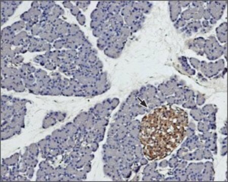
  • immunohistochemistry
  • immunoprecipitation from hippocampal lysate
  • immunohistochemical analysis

Monoclonal anti-syntaxin antibody can be used in immunoprecipitation and immunohistochemistry. It can also be used in western blotting. Further, this product can also be used to label monolayer cultures of neonatal retinal cells and frozen paraformaldehyde fixed tissue sections.

Biochem/physiol Actions

Syntaxin 1 (STX1A) may control exocytosis in synaptic vesicles. In retina, STX1A may help in the structural formation of the inner nuclear layer (INL) and outer plexiform layer (OPL). Deletion of STX1A in mice affects the functions of hypothalamic-pituitary-adrenal axis. It may participate in spinal nociceptive plasticity promoted by peripheral nerve injury. It has been implicated in docking at synaptic vesicles of presynaptic neurotransmitter release sites.

Disclaimer

Unless otherwise stated in our catalog or other company documentation accompanying the product(s), our products are intended for research use only and are not to be used for any other purpose, which includes but is not limited to, unauthorized commercial uses, in vitro diagnostic uses, ex vivo or in vivo therapeutic uses or any type of consumption or application to humans or animals.

Not finding the right product?  

Try our Product Selector Tool.

Storage Class Code

10 - Combustible liquids

WGK

WGK 3

Flash Point(F)

Not applicable

Flash Point(C)

Not applicable


Certificates of Analysis (COA)

Search for Certificates of Analysis (COA) by entering the products Lot/Batch Number. Lot and Batch Numbers can be found on a product’s label following the words ‘Lot’ or ‘Batch’.

Already Own This Product?

Find documentation for the products that you have recently purchased in the Document Library.

Visit the Document Library

M Tagaya et al.
The Journal of biological chemistry, 270(27), 15930-15933 (1995-07-07)
Syntaxin 1 (HPC-1), a component of the receptor for SNAPs (soluble N-ethylmaleimide-sensitive factor attachment proteins), has been implicated in the docking and fusion of synaptic vesicles with the plasma membrane. It was reported that syntaxin 1 in rat brain and
Shengxiu Li et al.
Investigative ophthalmology & visual science, 47(5), 2141-2149 (2006-04-28)
To examine the expression and cellular distribution pattern of endothelial nitric oxide synthase (eNOS) in the developing human retina and to compare its expression with that in rats. Expression of eNOS was examined by immunohistochemistry in retinas of humans ranging
Neuron-like differentiation and selective ablation of undifferentiated embryonic stem cells containing suicide gene with Oct-4 promoter
Hara A, et al.
Stem Cells and Development, 17(4), 619-627 (2008)
W Zhang et al.
The Journal of biological chemistry, 275(52), 41521-41527 (2000-10-12)
The Munc-18 protein (mammalian homologue of the unc-18 gene; also called nSec1 or rbSec1) has been identified as an essential component of the synaptic vesicle fusion protein complex. The cellular and subcellular localization and functional role of Munc-18 protein in
Stephanie L Fowler et al.
Cell communication & adhesion, 16(5-6), 117-130 (2009-10-23)
Gap junction proteins are a highly homologous family of 21 connexins. Here, the authors describe a tissue-specific technical artifact complicating analysis of connexin32 protein expression in the central nervous system. The authors show that in brain, but not liver, eight

Our team of scientists has experience in all areas of research including Life Science, Material Science, Chemical Synthesis, Chromatography, Analytical and many others.

Contact Technical Service