Skip to Content
Merck
All Photos(3)

Key Documents

SAB3500343

Sigma-Aldrich

Anti-Bax antibody produced in rabbit

IgG fraction of antiserum, buffered aqueous solution

Synonym(s):

Anti-Bax

Sign Into View Organizational & Contract Pricing


About This Item

MDL number:
UNSPSC Code:
12352203
NACRES:
NA.41

biological source

rabbit

Quality Level

conjugate

unconjugated

antibody form

IgG fraction of antiserum

antibody product type

primary antibodies

clone

polyclonal

form

buffered aqueous solution

species reactivity

human

technique(s)

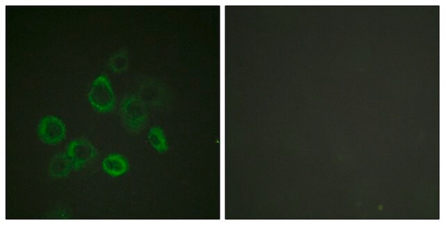
immunocytochemistry: suitable
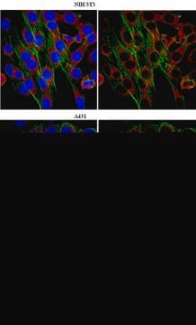
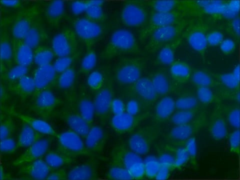
immunofluorescence: suitable
immunoprecipitation (IP): suitable
indirect ELISA: suitable
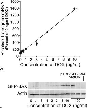
western blot: suitable

NCBI accession no.

UniProt accession no.

shipped in

dry ice

storage temp.

−20°C

target post-translational modification

unmodified

Gene Information

human ... BAX(581)

General description

BCL2 associated X, apoptosis regulator (BAX) is a proapoptotic protein of the B-cell lymphoma 2 (BCL2) protein family. The gene encoding it is localized on human chromosome 19q13.33.

Immunogen

Bax antibody was raised against a peptide corresponding to 16 amino acids near the amino-terminus of human Bax.

Application

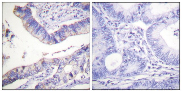
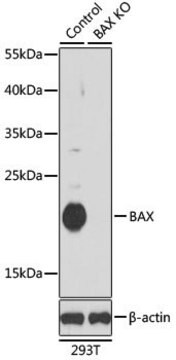
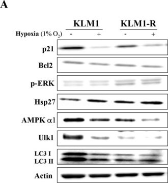
Anti-Bax antibody produced in rabbit has been used in Western blotting.

Biochem/physiol Actions

BCL2 associated X, apoptosis regulator (BAX) forms a heterodimer with B-cell lymphoma 2 (BCL2) and functions as an apoptotic activator. BAX interacts with and increases the opening of the mitochondrial voltage-dependent anion channel (VDAC), which leads to the loss in the mitochondrial membrane potential and the release of cytochrome c. The expression of BAX gene is regulated by the tumor suppressor p53 and BAX has been shown to be involved in p53-mediated apoptosis.

Features and Benefits

Evaluate our antibodies with complete peace of mind. If the antibody does not perform in your application, we will issue a full credit or replacement antibody. Learn more.

Linkage

The action of this antibody can be blocked using blocking peptide SBP3500343.

Physical form

Supplied in PBS with 0.02% sodium azide.

Disclaimer

Unless otherwise stated in our catalog or other company documentation accompanying the product(s), our products are intended for research use only and are not to be used for any other purpose, which includes but is not limited to, unauthorized commercial uses, in vitro diagnostic uses, ex vivo or in vivo therapeutic uses or any type of consumption or application to humans or animals.

Not finding the right product?  

Try our Product Selector Tool.

related product

Product No.
Description
Pricing

Storage Class Code

10 - Combustible liquids

WGK

WGK 2

Flash Point(F)

Not applicable

Flash Point(C)

Not applicable


Choose from one of the most recent versions:

Certificates of Analysis (COA)

Lot/Batch Number

Don't see the Right Version?

If you require a particular version, you can look up a specific certificate by the Lot or Batch number.

Already Own This Product?

Find documentation for the products that you have recently purchased in the Document Library.

Visit the Document Library

Wnt signal transduction pathway and apoptosis: a review
Nives Pecina-Slaus
Cancer Cell International (2010)
HspB8 mediates neuroprotection against OGD/R in N2A cells through the phosphoinositide 3-kinase/Akt pathway
Zhiping Hu
Brain Research (2016)
Bcl-2 family proteins and cancer
K W Yip
Oncogene (2008)
Jiajun Chen et al.
Experimental and therapeutic medicine, 23(6), 435-435 (2022-05-25)
Unveiling the mechanism of miR-122-5p in the mediation of forkhead box O3 (FOXO3) in regards to cochlear hair cell damage provides an effective solution for the treatment of ear hearing disorders. An oxidative stress model using a mouse cochlear hair
The role of Bcl-2 and its pro-survival relatives in tumourigenesis and cancer therapy
P N Kelly
Cell Death and Differentiation (2011)

Our team of scientists has experience in all areas of research including Life Science, Material Science, Chemical Synthesis, Chromatography, Analytical and many others.

Contact Technical Service