Skip to Content
Merck
All Photos(7)

Key Documents

T2200

Sigma-Aldrich

Anti-β-Tubulin III Antibody

enhanced validation

rabbit polyclonal

Synonym(s):

Anti-Tuj 1

Sign Into View Organizational & Contract Pricing


About This Item

MDL number:
UNSPSC Code:
12352203
NACRES:
NA.41

product name

Anti-β-Tubulin III antibody produced in rabbit, affinity isolated antibody, buffered aqueous solution

biological source

rabbit

Quality Level

conjugate

unconjugated

antibody form

affinity isolated antibody

antibody product type

primary antibodies

clone

polyclonal

form

buffered aqueous solution

mol wt

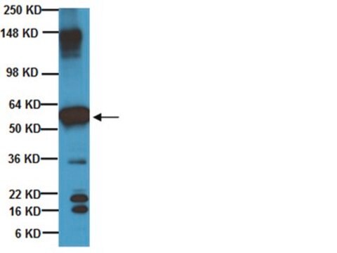
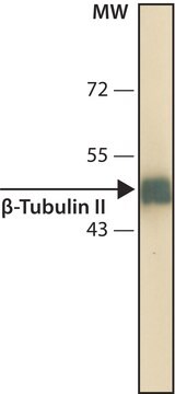
antigen ~55 kDa

species reactivity

rat, human, mouse

enhanced validation

knockout
Learn more about Antibody Enhanced Validation

technique(s)

immunoprecipitation (IP): 10 μg using RIPA extract (250 μg) of cultured rat PC12 cells
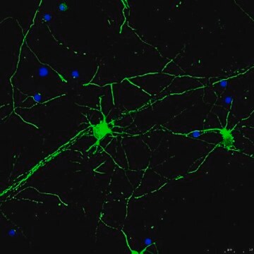
indirect immunofluorescence: 10-20 μg/mL using rat PC12 cells
western blot: 0.2-0.4 μg/mL using whole extracts of mouse brain or cultured human neuroblastoma SH-SY5Y cells

UniProt accession no.

shipped in

dry ice

storage temp.

−20°C

target post-translational modification

unmodified

Gene Information

human ... TUBB3(10381)
mouse ... Tubb3(22152)
rat ... Tubb3(246118)

Looking for similar products? Visit Product Comparison Guide

General description

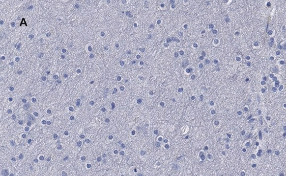
Tubulin β 3 class III (TUBB3) also known as β-Tubulin III, is encoded by the gene mapped to human chromosome 16q24.3. TUBB3 protein expression is restricted to neurons.
α/β-Tubulin, an integral component of microtubules, is present in almost all eukaryotic cells.
α/β-Tubulin occurs mostly as soluble (approx. 100-110 kDa) heterodimeric sets of α- and β-tubulin isotypes or as polymers in assembled microtubules. β-Tubulin III (also designated β-4 chain) is found in the brain and dorsal root ganglia and appears to be localized to neurons of the central and peripheral nervous system. β-tubulin III is also found in Sertoli cells of the testis, spermatozoa tails, certain lung cells, and apparently some breast stromal cells.

Specificity

Anti-β-Tubulin III recognizes human, mouse,and rat β-tubulin III.

Immunogen

synthetic peptide corresponding to amino acid residues 441-450 of human β-tubulin III (Ala446 to Ser446 substitution) with N-terminal added cysteine, conjugated to KLH. The sequence is conserved in mammals.

Application

Anti-β-Tubulin III antibody produced in rabbit has been used in following studies:
  • Immunofluorescence
  • Immunostaining
  • Western blotting
Anti-β-Tubulin III antibody produced in rabbit has been used in following studies:
  • immunofluorescence
  • immunostaining
  • western blotting
  • indirect immunoperoxidase assay (IPA)

Biochem/physiol Actions

Tubulin β 3 class III (TUBB3) plays a vital role in nervous system development and axon guidance. Mutation of the gene encoding protein leads to the ocular motility disorder, congenital fibrosis of the extraocular muscle type 3 (CFEOM3), and various neurological syndromes. Altered expression of the protein affects microtubule dynamics and microtubule-kinesin interactions. Increased expression of TUBB3 is observed in tumor tissues. Decreased expression of TUBB3 can be considered as an important marker for poor prognosis of cutaneous malignant melanoma. TUBB3 is implicated in the suppression of invasive growth of tumor tissue;therefore, it is considered to be a potential target for development of antitumor drugs.

Physical form

Solution in 0.01 M phosphate buffered saline, pH 7.4, containing 1% bovine serum albumin and 15 mM sodium azide.

Preparation Note

Storage and Stability: For continuous use, store at 2-8 °C for up to one month.
For extended storage, freeze in working aliquots. Repeated freezing and thawing is not recommended. Storage in frost-free freezers is also not recommended. If slight turbidity occurs upon prolonged storage, clarify the solution by centrifugation before use. Working dilutions should be discarded if not used within 12 hours.

Other Notes

In order to obtain the best results using various techniques and preparations, we recommend determining the optimal working concentrations by titration.

Disclaimer

Unless otherwise stated in our catalog or other company documentation accompanying the product(s), our products are intended for research use only and are not to be used for any other purpose, which includes but is not limited to, unauthorized commercial uses, in vitro diagnostic uses, ex vivo or in vivo therapeutic uses or any type of consumption or application to humans or animals.

Not finding the right product?  

Try our Product Selector Tool.

Storage Class Code

12 - Non Combustible Liquids

WGK

WGK 1

Flash Point(F)

Not applicable

Flash Point(C)

Not applicable


Certificates of Analysis (COA)

Search for Certificates of Analysis (COA) by entering the products Lot/Batch Number. Lot and Batch Numbers can be found on a product’s label following the words ‘Lot’ or ‘Batch’.

Already Own This Product?

Find documentation for the products that you have recently purchased in the Document Library.

Visit the Document Library

Characterization of the colchicine binding site on avian tubulin isotype βVI.
Sharma S, et al.
Biochemistry, 49(13), 2932-2942 (2010)
Geometric effect of cell adhesive polygonal micropatterns on neuritogenesis and axon guidance.
Jang MJ, Nam Y.
Journal of Neural Engineering, 9(4) (2012)
Immunohistochemical, volumetric, and functional neuroimaging studies in patients with idiopathic Parkinson's disease.
Hummel T, et al.
Journal of the Neurological Sciences, 289(1-2), 119-122 (2010)
Reciprocal regulation of the basic helix-loop-helix/Per-Arnt-Sim partner proteins, Arnt and Arnt2, during neuronal differentiation.
Hao N
Nucleic Acids Research, 41(11), 5626-5638 (2013)
C Gouarné et al.
British journal of pharmacology, 172(1), 235-245 (2014-09-16)
Parkinson's disease (PD) is usually diagnosed clinically from classical motor symptoms, while definitive diagnosis is made postmortem, based on the presence of Lewy bodies and nigral neuron cell loss. α-Synuclein (ASYN), the main protein component of Lewy bodies, clearly plays

Protocols

Cultivate ReNcell® human neural stem cells in 3D hydrogels for high-throughput screening using the TrueGel3D® HTS Hydrogel Plate with this protocol.

Our team of scientists has experience in all areas of research including Life Science, Material Science, Chemical Synthesis, Chromatography, Analytical and many others.

Contact Technical Service