Skip to Content
MilliporeSigma
All Photos(4)

Key Documents

L0668

Sigma-Aldrich

Anti-LAMP2 antibody produced in rabbit

affinity isolated antibody, buffered aqueous solution

Synonym(s):

Anti-CD107B, Anti-LGP110, Anti-Lysosomal Membrane Glycoprotein-type B (LGP-B), Anti-Lysosome-Associated Membrane Protein 2

Sign Into View Organizational & Contract Pricing


About This Item

MDL number:
UNSPSC Code:
12352203
NACRES:
NA.41

biological source

rabbit

Quality Level

conjugate

unconjugated

antibody form

affinity isolated antibody

antibody product type

primary antibodies

clone

polyclonal

form

buffered aqueous solution

mol wt

antigen ~110 kDa

species reactivity

human (predicted), mouse, rat

technique(s)

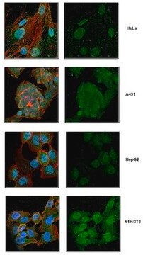
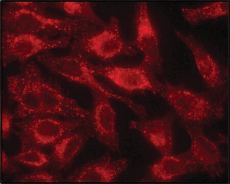
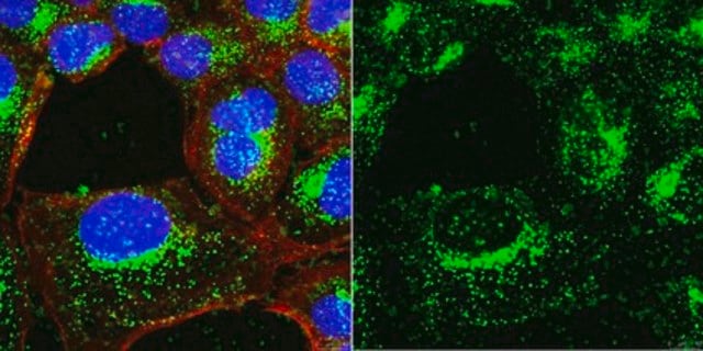
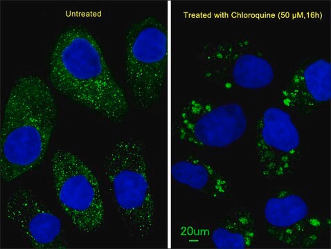
indirect immunofluorescence: 10-20 μg/mL using rat NRK and mouse NIH3T3 cells
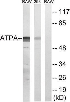
western blot (chemiluminescent): 2.5-5 μg using whole extract of rat NRK cells

UniProt accession no.

shipped in

dry ice

storage temp.

−20°C

target post-translational modification

unmodified

Gene Information

human ... LAMP2(3920)
mouse ... Lamp2(16784)
rat ... Lamp2(24944)

General description

LAMP2 (lysosome-associated membrane protein 2) is a heavily glycosylated intrinsic protein (110kD) present in lysosomal membrane. The protein glycosylation is essential for its function to protect the lysosomal membrane from proteolytic enzymes. Anti-LAMP2 antibody can be used in western blotting. It can also be used as a lysosomal marker. Rabbit anti-LAMP2 antibody reacts specifically with LAMP2 of mouse and rat.
Lysosomal associated membrane protein 2 (LAMP2) gene is located on human chromosome Xq24. LAMP2 is majorly expressed in lysosomes. It has three isoforms, that is LAMP2A, LAMP2B and LAMP2C.

Immunogen

synthetic peptide corresponding to amino acid residues 404-415 of mouse LAMP2 with N-terminal added cysteine, conjugated to KLH. The corresponding sequence is identical in rat and differs by two amino acids in human.

Application

Anti-LAMP2 antibody can be used in immunofluorescence and immunoblotting
Anti-LAMP2 antibody produced in rabbit has been used in immunohistochemical analysis.
Applications in which this antibody has been used successfully, and the associated peer-reviewed papers, are given below.
Western Blotting (1 paper)

Biochem/physiol Actions

Lysosomal associated membrane protein 2 (LAMP2) promotes cell-cell adhesion. It maintains cellular homeostasis and stress.

Target description

LAMP2 encodes a member of a family of membrane glycoproteins. This glycoprotein provides selectins with carbohydrate ligands. It may play a role in tumor cell metastasis. It may also function in the protection, maintenance, and adhesion of the lysosome.

Physical form

Solution in 0.01 M phosphate buffered saline, pH 7.4, containing 15 mM sodium azide.

Disclaimer

Unless otherwise stated in our catalog or other company documentation accompanying the product(s), our products are intended for research use only and are not to be used for any other purpose, which includes but is not limited to, unauthorized commercial uses, in vitro diagnostic uses, ex vivo or in vivo therapeutic uses or any type of consumption or application to humans or animals.

Not finding the right product?  

Try our Product Selector Tool.

Storage Class Code

10 - Combustible liquids

WGK

WGK 3

Flash Point(F)

Not applicable

Flash Point(C)

Not applicable

Personal Protective Equipment

dust mask type N95 (US), Eyeshields, Gloves

Choose from one of the most recent versions:

Certificates of Analysis (COA)

Lot/Batch Number

Don't see the Right Version?

If you require a particular version, you can look up a specific certificate by the Lot or Batch number.

Already Own This Product?

Find documentation for the products that you have recently purchased in the Document Library.

Visit the Document Library

Autoantibodies to hLAMP-2 in ANCA-negative pauci-immune focal necrotizing GN
Peschel A, et al.
Journal of the American Society of Nephrology, 25(3), 455-463 (2014)
Surface LAMP-2 is an endocytic receptor that diverts antigen internalized by human dendritic cells into highly immunogenic exosomes
Leone DA, et al.
Journal of Immunology, ji1601263-ji1601263 (2017)
Acute alloxan renal toxicity in the rat initially causes degeneration of thick ascending limbs of Henle
Terayama Y, et al.
Journal of Toxicologic Pathology (2016)
Ke-Qiong Deng et al.
Circulation, 137(14), 1486-1504 (2017-12-13)
Cardiac hypertrophy and its resultant heart failure are among the most common causes of mortality worldwide. Abnormal protein degradation, especially the impaired lysosomal degradation of large organelles and membrane proteins, is involved in the progression of cardiac hypertrophy. However, the
Olga S Sizova et al.
Cellular signalling, 26(4), 665-672 (2014-01-01)
We previously reported activation of the unfolded protein response (UPR) in P23H rhodopsin (RHO) retinas with autosomal dominant retinitis pigmentosa (ADRP). Knowing that the UPR can trigger Ca(2+) release from the endoplasmic reticulum and regulate cellular signaling we examined the

Our team of scientists has experience in all areas of research including Life Science, Material Science, Chemical Synthesis, Chromatography, Analytical and many others.

Contact Technical Service