Skip to Content
Merck
All Photos(1)

Key Documents

AB3612

Sigma-Aldrich

Anti-Caspase 12 Antibody, prodomain, aa 100-116

Chemicon®, from rabbit

Sign Into View Organizational & Contract Pricing


About This Item

UNSPSC Code:
12352203
eCl@ss:
32160702
NACRES:
NA.41

biological source

rabbit

Quality Level

antibody form

affinity purified immunoglobulin

antibody product type

primary antibodies

clone

polyclonal

purified by

affinity chromatography

species reactivity

human, rat, mouse

manufacturer/tradename

Chemicon®

technique(s)

immunohistochemistry: suitable
western blot: suitable

isotype

IgG

UniProt accession no.

shipped in

wet ice

target post-translational modification

unmodified

Gene Information

General description

Three distinct signaling pathways lead to apoptosis. The death receptor and mitochondrion are the two main pathways which involve caspase-8 and caspase-9, respectively. The endoplasmic reticulum stress is the third apoptotic pathway and involves caspase-12. Localized to the ER but not to cytoplasm or mitochondria, caspase-12 is activated by ER stress, including disruption of ER calcium homeostasis. Caspase-12 is co-localized to the ER with several proteins that are involved in Alzheimer′s disease including γ-secretase presenilin and ß-amyloid precursor protein (APP). Caspase-12 mediates ER stress-induced apoptosis as well as cytotoxicity induced by amyloid-ß. Caspase-12 is ubiquitously expressed in mouse tissues.

Specificity

Antibody recognizes caspase-12 pro-protein in mouse extracts of normal and apoptotic cells. Caspase-12 is predicted to have a large, >120 aa, long prodomain region. Thus this antibody recognizes only the proforms of the enzyme and not the active subunits. Uncleaved (proform) caspase-12 is prominently detected in many tissues. Active forms are difficult to detect expect in transfect cell systems. {Kalai, M et al (2003) 162:457-467}.

Immunogen

Epitope: prodomain, aa 100-116
Synthetic peptide corresponding to amino acids 100-116 of murine caspase-12.

Application

Detect Caspase 12 using this Anti-Caspase 12 Antibody, prodomain, aa 100-116 validated for use in WB, IH.
Research Category
Apoptosis & Cancer

Metabolism
Research Sub Category
Caspases

Enzymes & Biochemistry
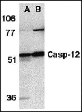
Western blot: 0.5 μg - 1 μg. Detects a band at ~49-53 kDa. in normal tissue extracts. Caspase-12 is a stress induced caspase localized to the ER and activated upon ER stress. Active process forms are difficult to detect in most non-transfected systems. Not all cells produce caspase-12; mouse L929 fibrosacrcoma cells are an excellent positive control line.

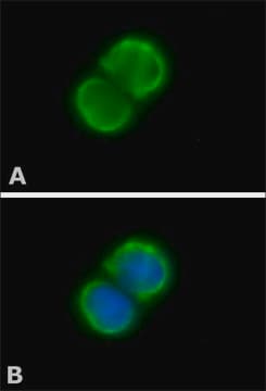
Immunohistochemistry: 2 μg /mL in mouse liver tissue

Mouse brain tissue lysate can be used as a positive control. Caspase 12 is associated with ER membranes and not in cytoplasm or mitochondria. Caspase 12 mediates cytotoxixity induced by amyloid beta.

Optimal working dilutions must be determined by end user.

Physical form

ImmunoAffinity Purified
Immunoaffinity chromatography purified antibody supplied in PBS containing 0.02% sodium azide.

Storage and Stability

Maintain at 2°-8°C for up to one year from date of receipt

Analysis Note

Control
Positive: mouse brain tissue lysate

Other Notes

Concentration: Please refer to the Certificate of Analysis for the lot-specific concentration.

Legal Information

CHEMICON is a registered trademark of Merck KGaA, Darmstadt, Germany

Disclaimer

Unless otherwise stated in our catalog or other company documentation accompanying the product(s), our products are intended for research use only and are not to be used for any other purpose, which includes but is not limited to, unauthorized commercial uses, in vitro diagnostic uses, ex vivo or in vivo therapeutic uses or any type of consumption or application to humans or animals.

Not finding the right product?  

Try our Product Selector Tool.

Storage Class Code

10 - Combustible liquids

WGK

WGK 2

Flash Point(F)

Not applicable

Flash Point(C)

Not applicable


Certificates of Analysis (COA)

Search for Certificates of Analysis (COA) by entering the products Lot/Batch Number. Lot and Batch Numbers can be found on a product’s label following the words ‘Lot’ or ‘Batch’.

Already Own This Product?

Find documentation for the products that you have recently purchased in the Document Library.

Visit the Document Library

Endoplasmic reticulum stress-associated caspase 12 mediates cisplatin-induced LLC-PK1 cell apoptosis.
Liu, H; Baliga, R
Journal of the American Society of Nephrology null
Liping Yang et al.
Neural regeneration research, 8(33), 3148-3158 (2014-09-11)
Endoplasmic reticulum stress is closely involved in the early stage of diabetic retinopathy. In the present study, a streptozotocin-induced diabetic animal model was given an intraperitoneal injection of tauroursodeoxycholic acid. Results from immunofluorescent co-localization experiments showed that both caspase-12 protein
Smriti A Agrawal et al.
Human molecular genetics, 26(14), 2667-2677 (2017-05-06)
Retinitis pigmentosa (RP) is the most common form of inherited retinal dystrophy. We recently identified mutations in REEP6, which encodes the receptor expression enhancing protein 6, in several families with autosomal recessive RP. REEP6 is related to the REEP and
Hua Liu et al.
World journal of gastroenterology, 20(48), 18189-18198 (2015-01-07)
To investigate the role of caspase-12 and its downstream targets in carbon tetrachloride (CCl4)-induced hepatocyte apoptosis. The role of caspase-12 was determined by using caspase-12 knock-out ((-/-)) mice. CCl4 (300 μL/kg body weight) or vehicle (corn oil) was administered to

Our team of scientists has experience in all areas of research including Life Science, Material Science, Chemical Synthesis, Chromatography, Analytical and many others.

Contact Technical Service