Skip to Content
Merck
All Photos(1)

Key Documents

356M-1

Sigma-Aldrich

Cytokeratin 5 & 6 (D5 & 16B4) Mouse Monoclonal Antibody

Sign Into View Organizational & Contract Pricing


About This Item

UNSPSC Code:
12352203
NACRES:
NA.41

biological source

mouse

Quality Level

100
500

conjugate

unconjugated

antibody form

culture supernatant

antibody product type

primary antibodies

clone

D5 & 16B4, monoclonal

description

For In Vitro Diagnostic Use in Select Regions (See Chart)

form

buffered aqueous solution

species reactivity

human

packaging

vial of 0.1 mL concentrate (356M-14)
vial of 0.5 mL concentrate (356M-15)
bottle of 1.0 mL predilute (356M-17)
vial of 1.0 mL concentrate (356M-16)
bottle of 7.0 mL predilute (356M-18)

manufacturer/tradename

Cell Marque

technique(s)

immunohistochemistry (formalin-fixed, paraffin-embedded sections): 1:50-1:200

isotype

IgG1

control

mesothelioma

shipped in

wet ice

storage temp.

2-8°C

visualization

cytoplasmic

Gene Information

human ... KRT5(3852)

General description

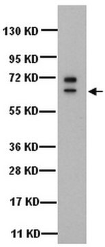
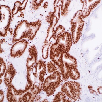
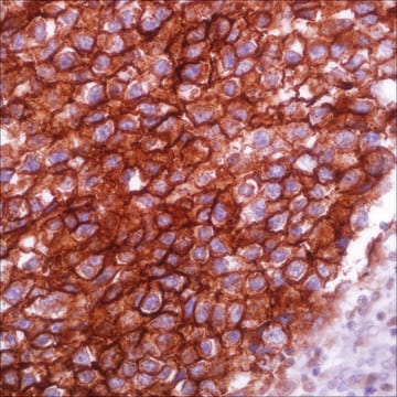
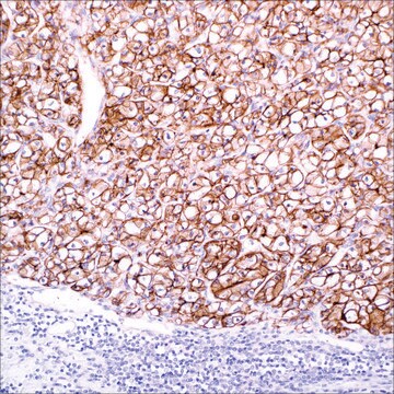
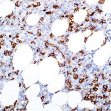
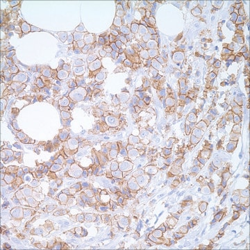
Anti-CK 5 & 6 positivity is seen in 65% to 100% of epithelioid mesotheliomas, in 0% to 19% of lung adenocarcinomas, and in 22% to 80% of serous carcinoma of the ovary and peritoneum. Keratin 5 & 6 expression in lung adenocarcinoma is not common, and when it occurs, the reaction is usually confined to small focal areas or scattered cells. The most probable cause of this finding is the presence of squamous differentiation of lung adenocarcinoma. Anti-CK 5 & 6 positivity has been useful in recognizing squamous cell carcinoma of the skin. Less than 10% of carcinomas of the breast, colon, and prostate stain positively for this marker. Anti-CK 5 & 6 has also been used successfully as a myoepithelial cell marker in the prostate and breast to determine malignancy. Anti-CK 5 & 6 is a useful marker to distinguish lung squamous cell carcinoma from lung adenocarcinoma and large cell carcinoma within a panel including antibodies against TTF-1, Napsin A, p63, SOX-2, desmocollin3, and desmoglein3.

Quality


IVD

IVD

IVD

RUO

Linkage

Cytokeratin 5 & 6 Positive Control Slides, Product No. 356S, are available for immunohistochemistry (formalin-fixed, paraffin-embedded sections).

Physical form

Solution in Tris Buffer, pH 7.3-7.7, with 1% BSA and <0.1% Sodium Azide

Preparation Note

Download the IFU specific to your product lot and formatNote: This requires a keycode which can be found on your packaging or product label.

Other Notes

For Technical Service please contact: 800-665-7284 or email: service@cellmarque.com

Legal Information

Cell Marque is a trademark of Merck KGaA, Darmstadt, Germany

Not finding the right product?  

Try our Product Selector Tool.


Certificates of Analysis (COA)

Search for Certificates of Analysis (COA) by entering the products Lot/Batch Number. Lot and Batch Numbers can be found on a product’s label following the words ‘Lot’ or ‘Batch’.

Already Own This Product?

Find documentation for the products that you have recently purchased in the Document Library.

Visit the Document Library

Magali Lacroix-Triki et al.
Virchows Archiv : an international journal of pathology, 442(6), 548-554 (2003-04-25)
Previous studies have shown that basal-type cytokeratins (CKs) can distinguish usual ductal hyperplasia (UDH) from the spectrum of atypical ductal hyperplasia (ADH), ductal carcinoma in situ (DCIS) and lobular carcinoma in situ (LCIS). Indeed, expression of these CKs is weak
Neil A Abrahams et al.
American journal of clinical pathology, 120(3), 368-376 (2003-09-25)
We evaluated the sensitivity and specificity of cytokeratin (CK) 5/6 for distinguishing foci of atrophy from prostatic adenocarcinoma with and without previous hormonal adjuvant therapy and observed the intensity and pattern of staining in mimickers of prostatic adenocarcinoma (basal cell
F Otterbach et al.
Histopathology, 37(3), 232-240 (2000-09-06)
This study was performed to determine the diagnostic value of keratin 5/6 (CK 5/6) immunophenotyping on routinely processed breast tissues. Six hundred and ninety-nine breast lesions, including normal tissues as well as benign and malignant lesions in 321 formalin-fixed, paraffin-embedded
J E Sigel et al.
Journal of cutaneous pathology, 28(10), 520-524 (2001-12-12)
Cutaneous spindle cell squamous cell carcinoma (SSCC) is a challenging diagnosis since it may be difficult to distinguish from spindle cell melanoma, leiomyosarcoma and atypical fibroxanthoma. Furthermore, it may be difficult to demonstrate epithelial differentiation by a traditional immunohistochemical panel.
Lin Lin et al.
Journal of cutaneous pathology, 30(2), 114-117 (2003-03-19)
Epithelioid sarcoma (ES) is a rare, aggressive soft tissue tumor characterized by nodular aggregates of epithelioid and/or spindled cells that are immunoreactive to cytokeratins (CKs) and epithelial membrane antigen. ES that arises in the dermis may cause epidermal ulceration and

Related Content

Diagnostic immunohistochemistry overview highlights its importance in cancer diagnosis and detecting infectious diseases in modern clinical pathology.

Our team of scientists has experience in all areas of research including Life Science, Material Science, Chemical Synthesis, Chromatography, Analytical and many others.

Contact Technical Service