Skip to Content
Merck
All Photos(6)

Key Documents

A5044

Sigma-Aldrich

Monoclonal Anti-α-Actinin antibody produced in mouse

clone BM-75.2, ascites fluid

Synonym(s):

Anti-BDPLT15

Sign Into View Organizational & Contract Pricing


About This Item

MDL number:
UNSPSC Code:
12352203
NACRES:
NA.41

biological source

mouse

Quality Level

conjugate

unconjugated

antibody form

ascites fluid

antibody product type

primary antibodies

clone

BM-75.2, monoclonal

mol wt

antigen 100 kDa

contains

15 mM sodium azide

species reactivity

mouse, bovine, human, chicken

technique(s)

indirect immunofluorescence: 1:200 using cultured chicken fibroblasts
microarray: suitable
western blot: suitable

isotype

IgM

UniProt accession no.

shipped in

dry ice

storage temp.

−20°C

target post-translational modification

unmodified

Gene Information

human ... ACTN1(87)
mouse ... Actn1(109711)

Related Categories

General description

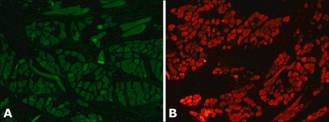
α-actinin is a 100kD actin binding protein found in muscle as well as non-muscle cells. In smooth muscle, α-actinin is identified in dense bodies and plaques characteristic of tissues whereas it is associated with z-discs that define muscle sarcomas in normal skeletal muscle. Monoclonal anti-α-actinin antibody can be used in immunoblotting to study the membrane anchorage sites and immunochemical identification of α-actinin. It can also be used in immunofluorescence to study the localization of α-actinin in cultured cells and tissues. Mouse anti-α-actinin antibody reacts specifically with chicken fibroblasts. The product has also shown reactivity for mouse and human cells.
The gene encoding α-actinin is mapped to the human chromosome 11q13.2. α-actinin belongs to the spectrin protein superfamily.

Immunogen

cytoskeletal fraction of bovine mammary gland epithelial (BMGE) cells.

Application

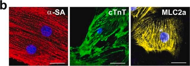
Immunofluorescence of cardiomyocytes from neonatal and adult mouse hearts was performed using monoclonal mouse anti-α actinin (clone EA-53) at 1:1000.
May be used in the study of skeletal muscle organization and in studies of the membrane anchorage sites of actin.
Monoclonal anti-α-actinin antibody (diluted 1: 500) can be used in native protein binding assays. It can also be used in western blot, microarray, immunohistochemistry and in Immunofluorescence microscopy.

Biochem/physiol Actions

α−actinin regulates muscle contraction and cytoskeletal organization. It promotes cell migration and acts as an actin crosslinker. Mutations in the α-actinin gene is observed in heterogeneous hypertrophic cardiomyopathy and in juvenile onset atrial fibrillation.

Disclaimer

Unless otherwise stated in our catalog or other company documentation accompanying the product(s), our products are intended for research use only and are not to be used for any other purpose, which includes but is not limited to, unauthorized commercial uses, in vitro diagnostic uses, ex vivo or in vivo therapeutic uses or any type of consumption or application to humans or animals.

Not finding the right product?  

Try our Product Selector Tool.

Storage Class Code

10 - Combustible liquids

WGK

nwg

Flash Point(F)

Not applicable

Flash Point(C)

Not applicable


Certificates of Analysis (COA)

Search for Certificates of Analysis (COA) by entering the products Lot/Batch Number. Lot and Batch Numbers can be found on a product’s label following the words ‘Lot’ or ‘Batch’.

Already Own This Product?

Find documentation for the products that you have recently purchased in the Document Library.

Visit the Document Library

C A Conley
American journal of physiology. Cell physiology, 280(6), C1645-C1656 (2001-05-15)
Evidence is accumulating to suggest that actin filament remodeling is critical for smooth muscle contraction, which implicates actin filament ends as important sites for regulation of contraction. Tropomodulin (Tmod) and smooth muscle leiomodin (SM-Lmod) have been found in many tissues
Protein Interactions with the Glucose Transporter Binding Protein GLUT1CBP That Provide a Link between GLUT1 and the Cytoskeleton.
Robert C. Bunn, Mari Anne Jensen
Molecular and Cellular Biology, 10(4), 819-832 (1999)
Thomas A Hawkins et al.
Development (Cambridge, England), 135(6), 1147-1156 (2008-02-08)
The mechanisms that regulate sarcomere assembly during myofibril formation are poorly understood. In this study, we characterise the zebrafish sloth(u45) mutant, in which the initial steps in sarcomere assembly take place, but thick filaments are absent and filamentous I-Z-I brushes
C M Laukaitis et al.
The Journal of cell biology, 153(7), 1427-1440 (2001-06-27)
To investigate the mechanisms by which adhesions form and disperse in migrating cells, we expressed alpha 5 integrin, alpha-actinin, and paxillin as green fluorescent protein (GFP) fusions. All localized with their endogenous counterparts and did not perturb migration when expressed
Zongwen Tian et al.
PloS one, 9(6), e100715-e100715 (2014-06-25)
Intracellular protein degradation is primarily performed by the ubiquitin-proteasome system (UPS) and the autophagic-lysosomal pathway (ALP). The interplay between these two pathways has been rarely examined in intact animals and the mechanism underlying the interplay remains unclear. Hence, we sought

Our team of scientists has experience in all areas of research including Life Science, Material Science, Chemical Synthesis, Chromatography, Analytical and many others.

Contact Technical Service