Skip to Content
Merck
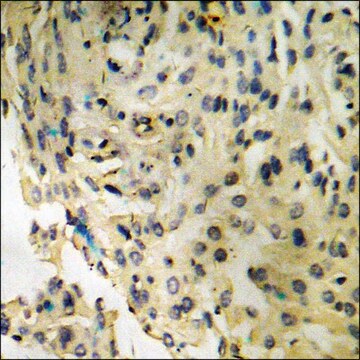
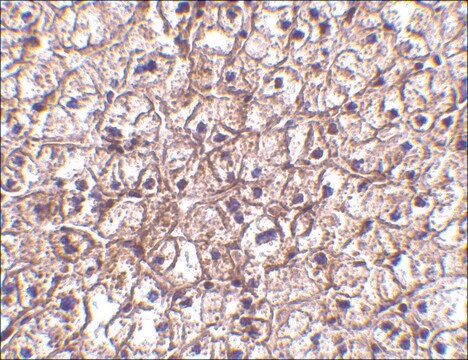
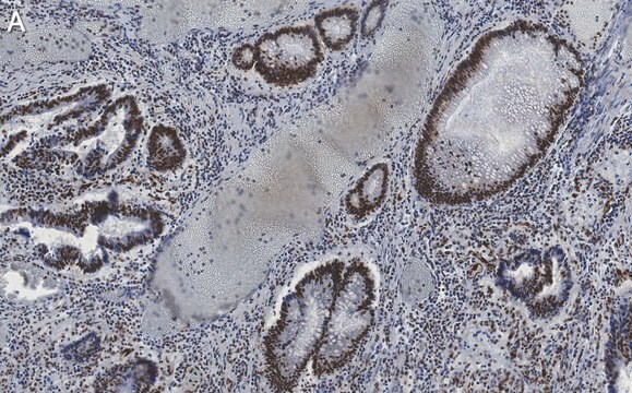
All Photos(4)

Key Documents

Safety Information

SAB4504031

Sigma-Aldrich

Anti-phospho-IRF-3 (pSer396) antibody produced in rabbit

affinity isolated antibody

Sign Into View Organizational & Contract Pricing


About This Item

UNSPSC Code:
12352203
NACRES:
NA.41

biological source

rabbit

Quality Level

conjugate

unconjugated

antibody form

affinity isolated antibody

antibody product type

primary antibodies

clone

polyclonal

form

buffered aqueous solution

mol wt

antigen 47 kDa

species reactivity

rat, mouse, human

concentration

~1 mg/mL

technique(s)

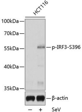
ELISA: 1:20000
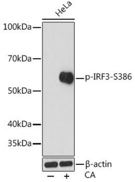
western blot: 1:500-1:1000

NCBI accession no.

UniProt accession no.

shipped in

wet ice

storage temp.

−20°C

target post-translational modification

phosphorylation (pSer396)

Gene Information

human ... IRF3(3661)

General description

IRF3 (interferon regulatory factor 3) is a transcription factor belonging to the IRF family and is involved in the innate immunity response against viruses and bacteria. The gene encoding IRF3 is localized on human chromosome 19q13.33.

Immunogen

The antiserum was produced against synthesized peptide derived from human IRF-3 around the phosphorylation site of Ser396.

Immunogen Range: 362-411

Application

Applications in which this antibody has been used successfully, and the associated peer-reviewed papers, are given below.
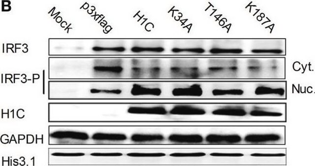
Western Blotting (1 paper)

Biochem/physiol Actions

Activated IRF3 (interferon regulatory factor 3) influences the expression of type I interferons via the toll-like receptor (TLR3/4)-mediated signaling pathway. IRF3 can suppress glioma invasion, proliferation and migration. Studies have suggested that IRF3 may have characteristics of tumor suppression.

Features and Benefits

Evaluate our antibodies with complete peace of mind. If the antibody does not perform in your application, we will issue a full credit or replacement antibody. Learn more.

Physical form

Rabbit IgG in phosphate buffered saline (without Mg2+ and Ca2+), pH 7.4, 150mM NaCl, 0.02% sodium azide and 50% glycerol.

Disclaimer

Unless otherwise stated in our catalog or other company documentation accompanying the product(s), our products are intended for research use only and are not to be used for any other purpose, which includes but is not limited to, unauthorized commercial uses, in vitro diagnostic uses, ex vivo or in vivo therapeutic uses or any type of consumption or application to humans or animals.

Not finding the right product?  

Try our Product Selector Tool.

Storage Class Code

10 - Combustible liquids

WGK

WGK 1

Flash Point(F)

Not applicable

Flash Point(C)

Not applicable


Regulatory Listings

Regulatory Listings are mainly provided for chemical products. Only limited information can be provided here for non-chemical products. No entry means none of the components are listed. It is the user’s obligation to ensure the safe and legal use of the product.

JAN Code

SAB4504031-100UG:


Certificates of Analysis (COA)

Search for Certificates of Analysis (COA) by entering the products Lot/Batch Number. Lot and Batch Numbers can be found on a product’s label following the words ‘Lot’ or ‘Batch’.

Already Own This Product?

Find documentation for the products that you have recently purchased in the Document Library.

Visit the Document Library

Interferon regulatory factor 3 alters glioma inflammatory and invasive properties
Leonid Tarassishin
Journal of Neuro-Oncology, 113 (2013)
Genetic susceptibility in cervical cancer: From bench to bedside
Afsane Bahrami
Journal of Cellular Physiology (2017)
Interferon regulatory factor 3 in adaptive immune responses.-Laure Ysebrant de Lendonck
Laure Ysebrant de Lendonck
Cellular and Molecular Life Sciences (2014)
Joyshree Karmakar et al.
Frontiers in immunology, 12, 626110-626110 (2021-03-26)
TLR4 activates two distinct signaling pathways involving adaptors MyD88 and TRIF to produce proinflammatory cytokines and type-I interferon respectively. How Leishmania donovani suppresses these pathways is not well studied. We earlier reported, TLR4 is hypersialylated due to reduced membrane-bound neuraminidase
Nikhil Jain et al.
Nature materials, 17(12), 1134-1144 (2018-10-24)
Macrophages respond to chemical/metabolic and physical stimuli, but their effects cannot be readily decoupled in vivo during pro-inflammatory activation. Here, we show that preventing macrophage spreading by spatial confinement, as imposed by micropatterning, microporous substrates or cell crowding, suppresses late

Our team of scientists has experience in all areas of research including Life Science, Material Science, Chemical Synthesis, Chromatography, Analytical and many others.

Contact Technical Service