
A1420
Ames′ Medium
With L-glutamine, without sodium bicarbonate, powder, suitable for cell culture
Select a Size
$620.00
Estimated to ship onMay 12, 2025
Select a Size
About This Item
$620.00
Estimated to ship onMay 12, 2025
Recommended Products


Quality Level
form
powder
technique(s)
cell culture | mammalian: suitable
components
glucose: 1.081 g/L (Dextro)
NaHCO3: no
L-glutamine: 0.073 g/L
shipped in
ambient
storage temp.
2-8°C
1 of 4
This Item | A1949 | A0545 | B3275 |
|---|---|---|---|
| species reactivity rabbit | species reactivity rabbit | species reactivity rabbit | species reactivity rabbit |
| biological source mouse | biological source mouse | biological source goat | biological source mouse |
| conjugate peroxidase conjugate | conjugate peroxidase conjugate | conjugate peroxidase conjugate | conjugate biotin conjugate |
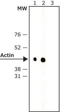
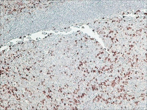
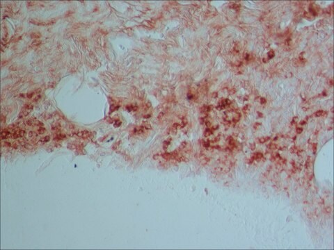
| technique(s) immunocytochemistry: suitable, western blot: 1:10,000-1:20,000 using 3T3 cell extracts using Anti-Actin (20-33), Catalog Number A5060, as the primary antibody, indirect ELISA: suitable, western blot: 1:10,000-1:20,000 using Hela cell extracts using Anti-Actin, N-terminal, Catalog Number A2103, as the primary antibody. | technique(s) direct ELISA: 1:40,000, immunohistochemistry (formalin-fixed, paraffin-embedded sections): 1:200, western blot: 1:160,000 using total cell extract of HeLa cells (5-10 μg/well) | technique(s) direct ELISA: 1:30,000 using using 5 μg/ml of rabbit IgG for the coating and OPD substrate, immunohistochemistry (formalin-fixed, paraffin-embedded sections): 1:200, western blot (chemiluminescent): 1:80,000-1:160,000 using detecting β-actin in total cell extract of HeLa cells (5-10 μg/mL) | technique(s) direct ELISA: 1:60,000, immunohistochemistry (formalin-fixed, paraffin-embedded sections): 1:1,500, western blot: 1:200,000-1:400,000 using using an assay detecting actin in total cell extract of HeLa cells (5-10 μg per well) |
| isotype IgG1 | isotype IgG1 | isotype - | isotype IgG1 |

General description
Application
Quantity
Reconstitution
Storage Class Code
11 - Combustible Solids
WGK
WGK 2
Flash Point(F)
Not applicable
Flash Point(C)
Not applicable
Choose from one of the most recent versions:
Certificates of Analysis (COA)
Don't see the Right Version?
If you require a particular version, you can look up a specific certificate by the Lot or Batch number.
Already Own This Product?
Find documentation for the products that you have recently purchased in the Document Library.
Customers Also Viewed

Our team of scientists has experience in all areas of research including Life Science, Material Science, Chemical Synthesis, Chromatography, Analytical and many others.
Contact Technical Service