Skip to Content
Merck
All Photos(1)

Key Documents

A276

Sigma-Aldrich

Monoclonal Anti-Na+/K+ ATPase (α Subunit) antibody produced in mouse

clone M7-PB-E9, ascites fluid

Synonym(s):

Anti-CMT2DD, Anti-HOMGSMR2

Sign Into View Organizational & Contract Pricing


About This Item

MDL number:
UNSPSC Code:
12352203
NACRES:
NA.41

biological source

mouse

Quality Level

conjugate

unconjugated

antibody form

ascites fluid

antibody product type

primary antibodies

clone

M7-PB-E9, monoclonal

mol wt

antigen ~110 kDa

species reactivity

canine, chicken, human, sheep, pig, bovine, mouse

should not react with

rat, Xenopus

technique(s)

immunocytochemistry: suitable
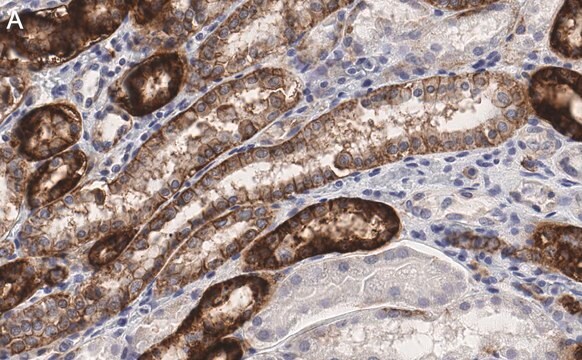
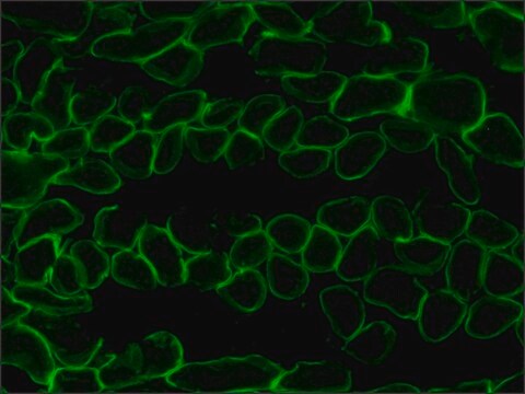
immunofluorescence: 1:20
immunohistochemistry (frozen sections): 1:100
immunoprecipitation (IP): suitable
indirect ELISA: suitable
western blot: 1:500-1:5000

isotype

IgG1

UniProt accession no.

shipped in

dry ice

storage temp.

−20°C

target post-translational modification

unmodified

Gene Information

human ... ATP1A1(476)

General description

The Na+/K+ ATPase (NKA) enzyme contains α, β and γ subunits. This enzyme is widely expressed. ATPase Na+/K+ transporting subunit α 1 (ATP1A1) is a principal catalytic subunit of NKA. It has a molecular mass of about 110kDa with 10 transmembrane segments. α-subunit has four different isoforms with tissue specific expression. α1 is primarily expressed in various tissues, including kidney, nerves and lung. While α2 is expressed in skeletal muscle and heart, α3 is found in the brain and α4 is apparently localized to testis and specifically to spermatozoa. ATP1A1 is encoded by the gene mapped to human chromosome 1p13-p11.

Immunogen

sheep kidney α subunit of Na+/K+ ATPase.

Application

Monoclonal Anti- Na+/K+ ATPase ( α Subunit) antibody produced in mouse has been used in flow cytometry.

Biochem/physiol Actions

The Na+/K+ ATPase is implicated in the maintenance of ion homeostasis for regular neuronal excitability, secondary transport and neurotransmitter uptake. ATPase Na+/K+ transporting subunit α 1 (ATP1A1) participates in both pumping and signaling functions. ATP1A1 regulates cellular transmembrane sodium and potassium ion gradients essential for the control of resting membrane potential. Additionally, nucleotide binding domain of ATP1A1 facilitates cellular signal transduction and cell growth. Deficiency of the gene leads to development of major depressive disorder (MDD).
The sodium/potassium ATPase is an integral membrane enzyme found in all cells of higher organisms and is responsible for the ATP-dependent transport of sodium and potassium across the cell membrane. This membrane-bound enzyme is related to a number of other ATPases including sarcoplasmic and endoplasmic reticulum calcium ATPase (SERCA) and plasma membrane calcium ATPase (PMCA). The sodium/potassium ATPase consists of a large, multipass, transmembrane catalytic subunit, termed the α subunit, and an associated smaller glycoprotein, termed the β subunit. Studies indicate that there are three isoforms of the α subunit (α 1, α 2, α 3) and two isoforms of the β subunit (β 1 and β 2) encoded by two multigene families.
The different isoforms of the sodium/potassium ATPase exhibit tissue-specific and developmental patterns of expression. The α 1 and β mRNAs are present in all cell types examined, whereas the α 2 and α 3 mRNAs exhibit a more restricted pattern of cell-specific expression. The α subunit has been found in kidney, brain, heart, and to a lesser extent liver, skeletal and smooth muscle.

Target description

Na+/K+ transporting ATPase subunit α1 is an ion transport pump critical for maintaining the gradient of Na and K ions across the plasma membrane.

Physical form

solution containing 1 mg/ml BSA and 0.05% sodium azide

Disclaimer

Unless otherwise stated in our catalog or other company documentation accompanying the product(s), our products are intended for research use only and are not to be used for any other purpose, which includes but is not limited to, unauthorized commercial uses, in vitro diagnostic uses, ex vivo or in vivo therapeutic uses or any type of consumption or application to humans or animals.

Not finding the right product?  

Try our Product Selector Tool.

Storage Class Code

13 - Non Combustible Solids

WGK

WGK 1

Flash Point(F)

Not applicable

Flash Point(C)

Not applicable

Personal Protective Equipment

dust mask type N95 (US), Eyeshields, Gloves

Certificates of Analysis (COA)

Search for Certificates of Analysis (COA) by entering the products Lot/Batch Number. Lot and Batch Numbers can be found on a product’s label following the words ‘Lot’ or ‘Batch’.

Already Own This Product?

Find documentation for the products that you have recently purchased in the Document Library.

Visit the Document Library

Jan Stindt et al.
Liver international : official journal of the International Association for the Study of the Liver, 33(10), 1527-1535 (2013-06-14)
The bile salt export pump (BSEP, ABCB11) is essential for bile salt secretion at the canalicular membrane of liver cells. Clinical phenotypes associated with BSEP mutations are commonly categorized as benign recurrent intrahepatic cholestasis (BRIC-2) or progressive familial intrahepatic cholestasis
Viviane Wilms et al.
Scientific reports, 6, 34203-34203 (2016-09-30)
In the cochlea, mammals maintain a uniquely high endolymphatic potential (EP), which is not observed in other vertebrate groups. However, a high [K+] is always present in the inner ear endolymph. Here, we show that Kir4.1, which is required in
Viviane Wilms et al.
The Journal of experimental biology, 220(Pt 15), 2701-2705 (2017-05-21)
Mechanoelectrical transduction in the vertebrate inner ear is a highly conserved mechanism that is dependent on K+ influx into hair cells. Here, we investigated the molecular underpinnings of subsequent K+ recycling in the chicken basilar papilla and compared them with
Interplay between a cytosolic and a cell surface carbonic anhydrase in pH homeostasis and acid tolerance of
Dhiman Sankar Pal et al.
Journal of cell science, 130(4), 754-766 (2017-01-08)
Eva Skiöldebrand et al.
Heliyon, 4(1), e00525-e00525 (2018-03-22)
Chondrocytes are effectively involved in the pathophysiological processes of inflammation in joints. They form cellular processes in the superficial layer of the articular cartilage and form gap junction coupled syncytium to facilitate cell-to-cell communication. However, very little is known about

Our team of scientists has experience in all areas of research including Life Science, Material Science, Chemical Synthesis, Chromatography, Analytical and many others.

Contact Technical Service