Skip to Content
MilliporeSigma
HomeBiopharmaceutical CharacterizationAnalysis of mAb and ADC on Widepore Silica Monolith Column

HPLC-UV Analysis of Native monoclonal antibody (mAb) and antibody-drug conjugate (ADC) on Widepore Silica Monolith Column Under High Flow Rate Conditions

Gisela Jung, R&D Scientist, Benjamin Peters, R&D Principal Scientist, Cory Muraco, Biomolecule Workflows Product Manager, Hillel Brandes, Analytical Technology Specialist

Introduction

 Monolithic silica columns for HPLC offer low backpressure due to their high porosity (> 80%). This aspect leads to the possibility of using relatively high flow rates, resulting in reduced analysis time.

In this work, the influence of different parameters like flow rate, temperature, and solvent additives is shown for the determination of intact universal antibody standard human (Figures 2-13) and antibody-drug conjugate (ADC) mimic (Figures 14-15) on a Chromolith® WP 300 RP-18 column. 
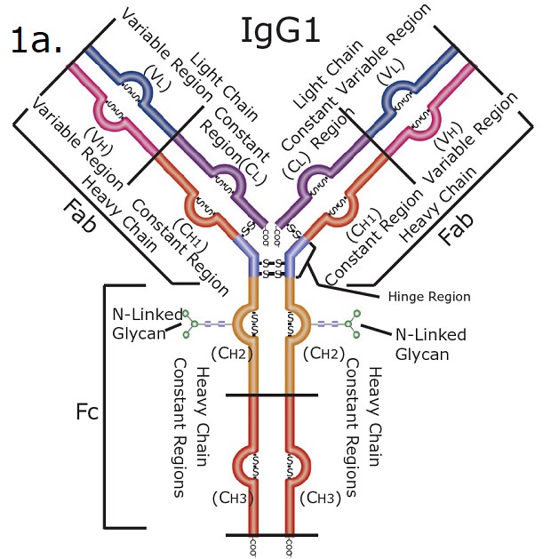
The structural diagram of SILu™Lite SigmaMAb Universal Antibody Standard human, highlighting its key regions and components. The antibody has a characteristic "Y" shape and is divided into distinct functional parts. The upper arms of the "Y" structure represent the Fab (fragment antigen-binding) regions. Each Fab contains a light chain and a heavy chain, with both having variable (VL and VH) and constant (CL and CH1) regions. The stem of the "Y" represents the Fc (fragment crystallizable) region, composed of constant domains of the heavy chains (CH2 and CH3). Located at the junction of the Fab and Fc regions, the hinge region provides flexibility to the antibody, allowing better antigen binding. N-Linked Glycans are shown attached to the CH2 domains in the Fc region. The glycan structures are depicted as branched molecules. Disulfide Bonds are depicted as "S-S" bridges, connecting the heavy and light chains as well as the two heavy chains.
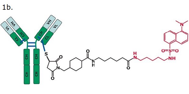
A structural representation of an antibody-drug conjugate (ADC) mimic. On the left side, the structure of an antibody is shown, with two heavy chains and two light chains. The heavy chains are depicted in green, with regions labeled CH (constant heavy) and VH (variable heavy), while the light chains are shown in grey, labeled CL (constant light) and VL (variable light). Disulfide bonds, represented as blue lines, connect the chains. Extending from the antibody is a linker-drug complex. The linker portion is depicted as a black chemical structure, connecting the antibody to the drug molecule. The drug portion is on the far right, displayed in red. It consists of a sulfonamide group attached to a bicyclic aromatic system, with additional functional groups, such as an amine and a methylated nitrogen, enhancing its distinct structure.

Figure 1. SILu™Lite SigmaMAb Universal Antibody Standard human (1a) and SigmaMAb Antibody-Drug Conjugate (ADC) Mimic (1b).
Experimental Method

SILu™Lite SigmaMAb standards (MSQC4) and SigmaMAb antibody drug conjugate mimic (MSQC8) standards.

The standard solutions were analyzed on a 100 x 2 mm I.D. Chromolith® WP 300 RP-18 column under conditions shown in Table 1.

Table 1.Conditions for analysis of SILuLite SigmaMAb by HPLC-UV

Results

Analysis of Universal Antibody Standard Human

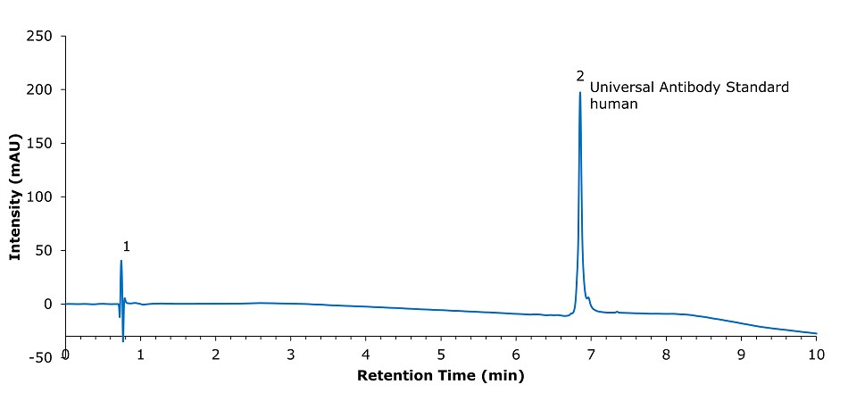
A chromatogram representing the analysis of a human antibody standard. The x-axis represents retention time in minutes, ranging from 0 to 10, while the y-axis represents intensity in milli-absorbance units (mAU), ranging from -50 to 250. Two peaks are observed. Peak 1, occurring near 0.7 minutes, is small and sharp, likely corresponding to unretained components or injection artifacts. Peak 2, appearing at approximately 7 minutes, is tall, sharp, and well-defined. It is labeled as "Universal Antibody Standard human," indicating the retention time of the primary analyte. The baseline remains stable throughout, with a gradual decline after Peak 2.

Figure 2.Analysis of SILu™Lite SigmaMAb Universal Antibody Standard human on a Chromolith® WP 300 RP-18 (100 x 2 mm I.D.).

Table 2.Chromatographic data for analysis of SigmaMAb Universal Antibody Standard human

Analysis of Universal Antibody Standard Human at Different Temperatures
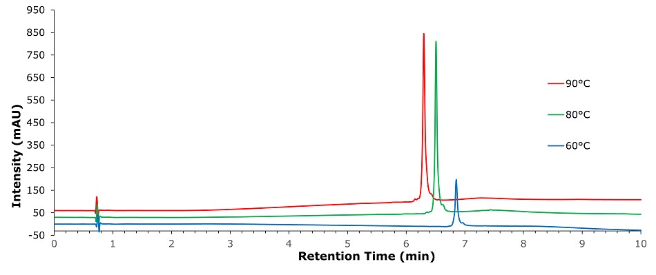
Overlayed chromatograms comparing the analysis of a human antibody standard on a Chromolith® WP 300 RP-18 (100 x 2 mm I.D.) column under different temperature conditions: 90°C (red line), 80°C (green line), and 60°C (blue line). The x-axis represents retention time in minutes, ranging from 0 to 10, while the y-axis represents intensity in milli-absorbance units (mAU), ranging from -50 to 900. Initial small peaks around 0.7 minutes for all temperatures, likely representing unretained components or injection artifacts. Main peaks appear between 6 and 7 minutes. The peak at 90°C (red) is the tallest, followed by 80°C (green) and 60°C (blue), indicating that higher temperatures improve signal intensity and resolution. Baseline stability varies slightly, with higher temperatures showing a slightly elevated baseline.

Figure 3.Analysis of SILu™Lite SigmaMAb Universal Antibody Standard human in matrix on a Chromolith® WP 300 RP-18 (100 x 2 mm I.D.) column at different temperatures

Table 3.Chromatographic data for analysis of SigmaMAb Universal Antibody Standard human at three different temperatures

Analysis of Universal Antibody Standard Human with Different Solvent Additives on a Chromolith® WP 300 RP-18 Column
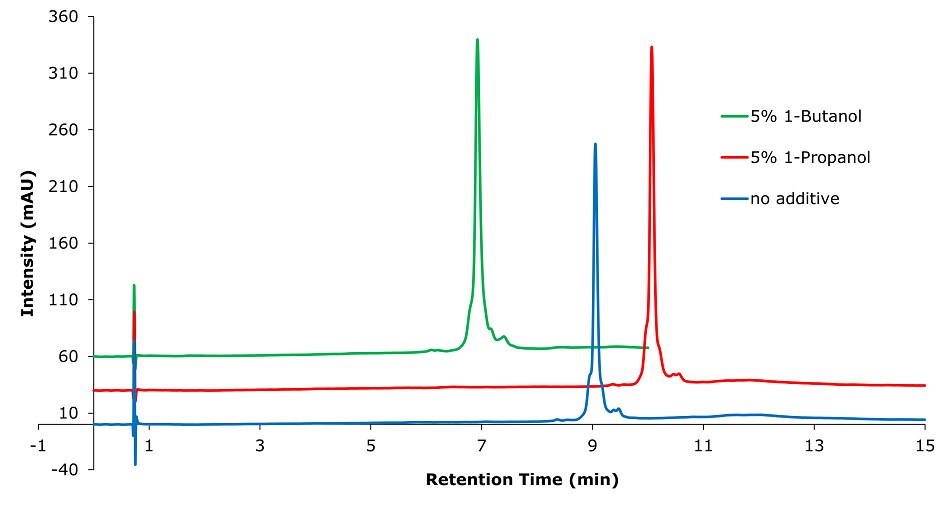
Overlayed chromatograms comparing the analysis of a human antibody standard on a Chromolith® WP 300 RP-18 (100 x 2 mm I.D.) column with different solvent additives: 5% 1-Butanol (green line), 5% 1-Propanol (red line), and no additive (blue line). The x-axis represents retention time in minutes, ranging from 0 to 15, while the y-axis represents intensity in milli-absorbance units (mAU), ranging from -10 to 360. Initial small peaks around 1 minute for all conditions, likely representing unretained components or injection artifacts. The main distinct peaks occur between 6 and 10 minutes. The green chromatogram (5% 1-Butanol) displays the tallest peak at around 7 minutes, followed by the red chromatogram (5% 1-Propanol) with its peak near 10 minutes, and the blue chromatogram (no additive) showing a smaller peak also around 9 minutes. Baseline stability varies, with the blue chromatogram (no additive) maintaining a lower baseline intensity compared to the others.

Figure 4.Analysis of SILu™Lite SigmaMAb Universal Antibody Standard human in matrix on a Chromolith® WP 300 RP-18 (100 x 2 mm I.D.) column with different solvent additives. Method: Temp. 60 °C, Mobile phase: A: 1-Propanol or 1-Butanol; B: Water 0.1% (v/v) TFA, C: Acetonitrile 0.1% (v/v) TFA; Gradient: 0 min A:5% B:70% C:25%, 0.1 min A:5% B:70% C:25%, 10 min A:5% B:60% C:35%.

Table 4.Chromatographic data for analysis of SigmaMAb Universal Antibody Standard human with different solvent additives on Chromolith® WP 300 RP-18 (100 x 2 mm I.D.) column

Analysis of Universal Antibody Standard Human with Different Solvent Additives on a BIOshell™ A400 Protein C18 Column
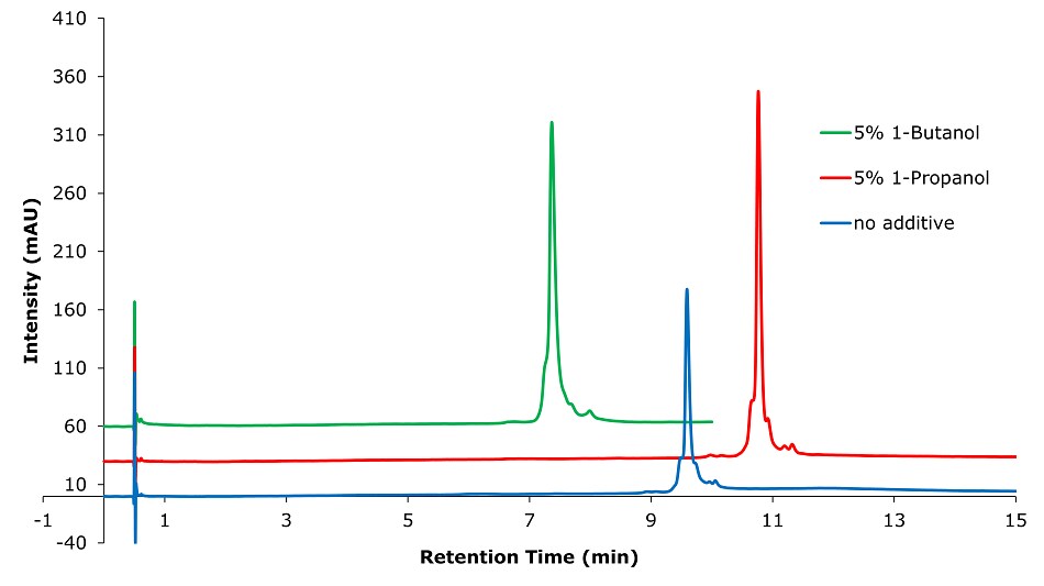
Overlayed chromatograms comparing the analysis of a human antibody standard on a BIOshellTM A400 Protein C18, 3.4 µm (100 x 2.1 mm I.D.) column with different solvent additives: 5% 1-Butanol (green line), 5% 1-Propanol (red line), and no additive (blue line). The x-axis represents retention time in minutes, ranging from 0 to 15, and the y-axis represents intensity in milli-absorbance units (mAU), ranging from -10 to 410. Initial peaks around 1 minute are small for all solvent conditions, representing unretained or early-eluting components. Main distinct peaks appear for the three chromatograms between 7 and 11 minutes. The green chromatogram (5% 1-Butanol) shows the highest peak intensity at around 7 minutes. The red chromatogram (5% 1-Propanol) displays a prominent peak near 11 minutes. The blue chromatogram (no additive) exhibits a smaller peak near 9 minutes with less intensity than the additive-containing conditions. Baseline stability varies, with the red and green chromatograms showing a slightly elevated baseline compared to the blue chromatogram.

Figure 5.Analysis of SILu™Lite SigmaMAb Universal Antibody Standard human in matrix on a BIOshell™ A400 Protein C18, 3.4 µm (100 x 2.1 mm I.D.) column with different solvent additives. Method: Temp. 60 °C, Mobile phase: A: 1-Propanol or 1-Butanol; B: Water 0.1% (v/v) TFA, C: Acetonitrile 0.1% (v/v) TFA; Gradient: 0 min A:5% B:70% C:25%, 0.1 min A:5% B:70% C:25%, 10 min A:5% B:60% C:35%.

Table 5.Chromatographic data for analysis of SigmaMAb Universal Antibody Standard human with different solvent additives on BIOshell™ A400 Protein C18, 3.4 µm (100 x 2 mm I.D.) column

Analysis of Universal Antibody Standard Human with Premixed Mobile Phase Compared to Mobile Phase Mixed by Instrument Pump on a Chromolith® WP 300 RP-18 Column
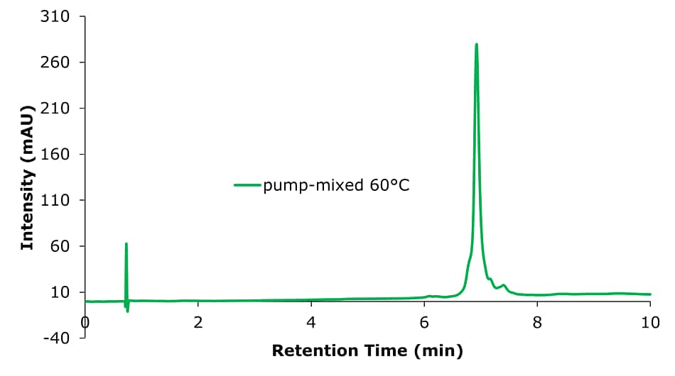
A chromatogram representing the analysis of a human antibody standard on a Chromolith® WP 300 RP-18 (100 x 2 mm I.D.) column with the mobile phase mixed by the instrument pump at 60°C. The x-axis represents retention time in minutes, ranging from 0 to 10, and the y-axis represents intensity in milli-absorbance units (mAU), ranging from -40 to 310. Initial peak near 1 minute is small and sharp, likely representing unretained components or injection artifacts. The main peak appears at approximately 7 minutes, exhibiting a tall, sharp, and well-defined profile, indicating the retention time of the antibody standard. This peak dominates the chromatogram, highlighting the efficient separation and detection of the analyte. The baseline remains stable and flat throughout the run, with no significant noise or artifacts.

Figure 6.Analysis of SILu™Lite SigmaMAb Universal Antibody Standard human on a Chromolith® WP 300 RP-18 (100 x 2 mm I.D.) column with mobile phase mixed by instrument pump. Method: Temp. 60 °C, Mobile phase: A: Water 0.1 % TFA; B: Acetonitrile 0.1% TFA, C: 1-Butanol; Gradient: 0 min A:70% B:25% C:5%, 0.1 min A:70% B:35% C:5%, 10 min A:60% B:35% C:5%.
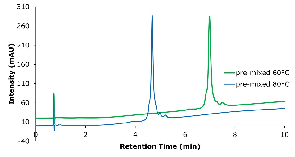
An overlay of chromatograms comparing the analysis of a human antibody standard using a pre-mixed mobile phase at two temperatures: 60°C (green line) and 80°C (blue line). The x-axis represents retention time in minutes, ranging from 0 to 10, and the y-axis represents intensity in milli-absorbance units (mAU), ranging from -40 to 310. Initial peaks around 1 minute are small and sharp for both conditions, likely representing unretained components or injection artifacts. At 80°C (blue line), a distinct tall  peak appears earlier, around 5 minutes, with a slightly higher intensity. At 60°C (green line), the peak appears later, around 6.5 minutes, with a broader profile but slightly lower intensity. Baseline behavior differs: the blue chromatogram (80°C) exhibits a lower baseline across the run, while the green chromatogram (60°C) shows a slight baseline rise.

Figure 7.Analysis of SILu™Lite SigmaMAb Universal Antibody Standard human on a Chromolith® WP 300 RP-18 (100 x 2 mm I.D.) column with pre-mixed mobile phase at two different temperatures. Mobile phase: A: Acetonitrile/ 1-Butanol/ Water 25/5/70 0.1% (v/v) TFA, B: Acetonitrile/ 1-Butanol/ Water 35/5/60 0.1% (v/v) TFA, Gradient: 0 min A:100% B:0%, 0.1 min A:100% B:0%, 10 min A:0% B:100%.

Table 6.Chromatographic data for analysis of SigmaMAb Universal Antibody Standard human on a Chromolith® WP 300 RP-18 (100 x 2 mm I.D.) column with pre-mixed mobile phase at two different temperatures and pump-mixed mobile phase

Analysis of Universal Antibody Standard Human with Different Flow Rates on a Chromolith® WP 300 RP-18 Column
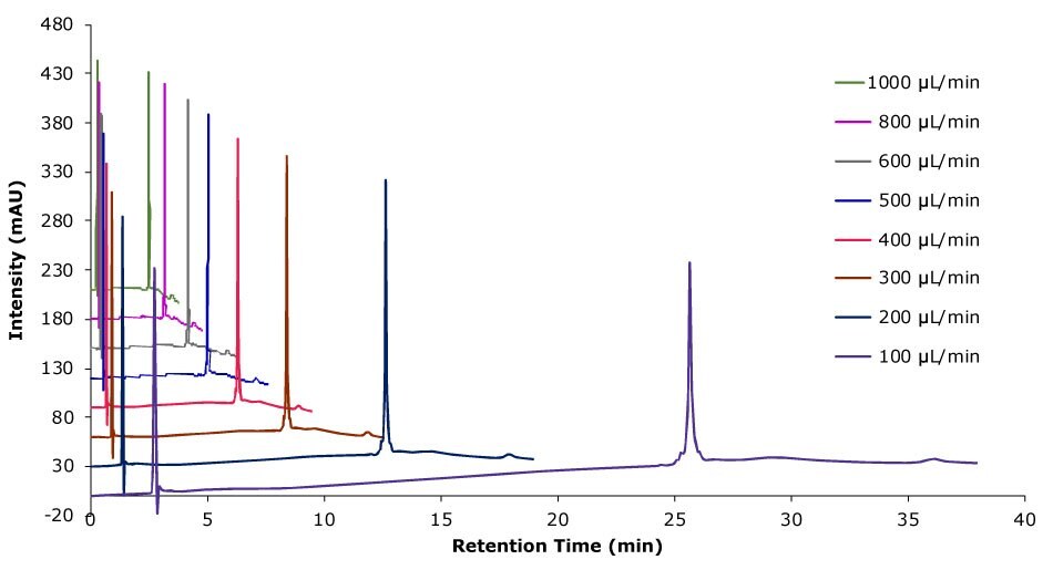
An overlayed chromatogram illustrating the impact of different flow rates on the analysis of SILu™Lite SigmaMAb Universal Antibody Standard human on a Chromolith® WP 300 RP-18 (100 x 2 mm I.D.) column with different flow rates. The x-axis represents retention time in minutes, ranging from 0 to 40, while the y-axis represents intensity in milli-absorbance units (mAU), ranging from -20 to 480. The chromatograms are color-coded to represent various flow rates: 1000 µL/min (red), 800 µL/min (yellow), 600 µL/min (mint-green), 500 µL/min (dark green), 400 µL/min (sky-blue), 300 µL/min (blue), 200 µL/min (dark blue), and 100 µL/min (purple). As the flow rate decreases the retention time for the antibody increases. At higher flow rates (e.g., 1000 µL/min and 800 µL/min), retention times are shorter, and the peaks are sharper and occur earlier, while at lower flow rates (e.g., 100 µL/min and 200 µL/min), retention times are longer, and peaks appear broader.

Figure 8.Analysis of SILu™Lite SigmaMAb Universal Antibody Standard human on a Chromolith® WP 300 RP-18 (100 x 2 mm I.D.) column with different flow rates. Method: Temp. 60 °C, Mobile phase: A: Water 0.1 % (v/v) TFA, B: Acetonitrile 0.1 % (v/v) TFA; Gradient: 20% B to 60% B, adjustment see table.
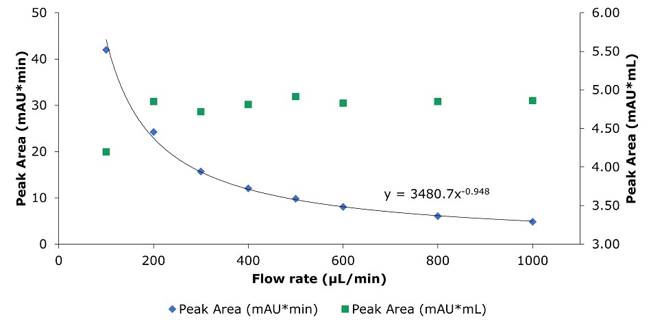
Table 7.Chromatographic data for analysis of SigmaMAb Universal Antibody Standard human on a Chromolith® WP 300 RP-18 (100 x 2 mm I.D.) column with different flow rates
A dual-axis graph illustrating the relationship between flow rate and peak area for SILu™Lite SigmaMAb Universal Antibody Standard human on a Chromolith® WP 300 RP-18 (100 x 2 mm I.D.) column. The x-axis represents flow rate in microliters per minute (µL/min), ranging from 0 to 1000. The left y-axis indicates peak area in milli-absorbance units multiplied by minutes (mAUmin), while the right y-axis represents peak area in milli-absorbance units multiplied by milliliters (mAUmL). Two sets of data points are displayed: blue diamonds for peak area in mAUmin and green squares for peak area in mAUmL. The blue diamonds follow a decreasing trend as flow rate increases, showing a curve fitted to the equation 𝑦=3480.7𝑥−0.948, which demonstrates an inverse relationship between flow rate and peak area in mAUmin. In contrast, the green squares remain relatively constant across the range of flow rates, indicating that the peak area in mAUmL is unaffected by changes in flow rate.

Figure 9.Analysis of SILu™Lite SigmaMAb Universal Antibody Standard human on a Chromolith® WP 300 RP-18 (100 x 2 mm I.D.) column. Peak area as a function of flow rate.
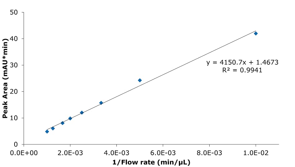
A scatter plot illustrating the relationship between peak area and the reciprocal of flow rate for the analysis of SILu™Lite SigmaMAb Universal Antibody Standard human on a Chromolith® WP 300 RP-18 (100 x 2 mm I.D.) column. The x-axis represents 1/Flow rate in min/μL, while the y-axis represents peak area in milli-absorbance units multiplied by minutes (mAU*min), ranging from 0 to 50. The data points, represented by blue diamonds, show a positive linear relationship, with peak area increasing as 1/Flow rate increases. A fitted linear regression line is shown, described by the equation y=4150.7x+1.4673, with an R2 value of 0.9941, indicating a very strong correlation. The graph highlights the dependency of peak area on the reciprocal of flow rate, demonstrating a predictable linear trend in the data.

Figure 10.Analysis of SILu™Lite SigmaMAb Universal Antibody Standard human on a Chromolith® WP 300 RP-18 (100 x 2 mm I.D.) column. Peak area as a function of 1/flow rate.
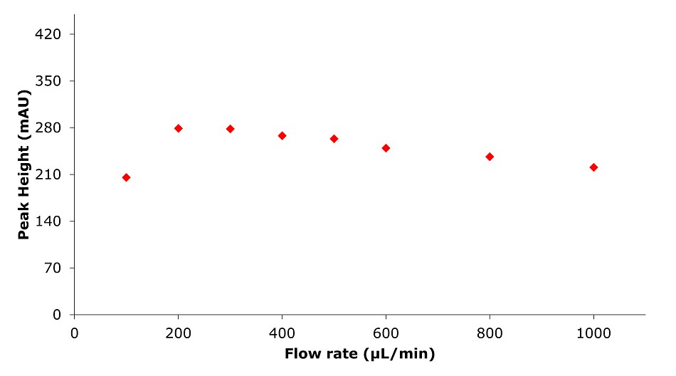
A scatter plot depicting the relationship between flow rate and peak height for the analysis of of SILu™Lite SigmaMAb Universal Antibody Standard human on a Chromolith® WP 300 RP-18 (100 x 2 mm I.D.) column. The x-axis represents flow rate in microliters per minute (µL/min), ranging from 0 to 1000, while the y-axis represents peak height in milli-absorbance units (mAU), ranging from 0 to 420. Data points, represented by red diamonds, show an initial increase in peak height with rising flow rate, reaching a plateau between 200 µL/min and 600 µL/min. Beyond 600 µL/min, the peak height begins to decrease slightly but remains relatively consistent. The plot indicates that peak height stabilizes at intermediate flow rates, demonstrating the optimal range for achieving maximal signal intensity during chromatographic analysis

Figure 11.Analysis of SILu™Lite SigmaMAb Universal Antibody Standard human on a Chromolith® WP 300 RP-18 (100 x 2 mm I.D.) column. Peak height as function of flow rate.
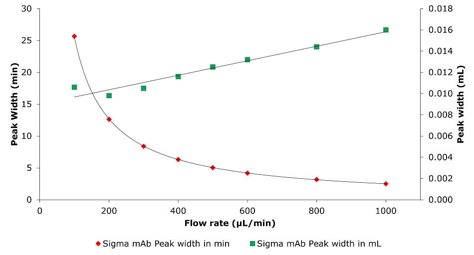
A dual-axis graph depicting the relationship between flow rate and peak width for the analysis of SILu™Lite SigmaMAb Universal Antibody Standard human on a Chromolith® WP 300 RP-18 (100 x 2 mm I.D.) column. The x-axis represents flow rate in microliters per minute (µL/min), ranging from 0 to 1000. The left y-axis represents peak width in minutes, while the right y-axis represents peak width in milliliters (mL). Two sets of data points are shown: red diamonds representing peak width in minutes and green squares representing peak width in milliliters. The red diamonds show a decreasing trend, with peak width in minutes decreasing significantly as flow rate increases, following a curved pattern. Conversely, the green squares show an increasing trend, with peak width in milliliters growing linearly with flow rate. This graph highlights the opposing trends in peak width depending on the unit of measurement, demonstrating the influence of flow rate on chromatographic peak characteristics.

Figure 12.Analysis of SILu™Lite SigmaMAb Universal Antibody Standard human on a Chromolith® WP 300 RP-18 (100 x 2 mm I.D.) column. Peak width as function of flow rate.

Analysis of ADC Mimic at Different Temperatures and with Additive

an overlayed chromatogram comparing the analysis of SILu™Lite ADC Mimic on a Chromolith® WP 300 RP-18 (100 x 2 mm I.D.) column at various temperatures and with a solvent additive. The x-axis represents retention time in minutes, ranging from 0 to 10, while the y-axis represents intensity in milli-absorbance units (mAU), ranging from -50 to 850. Four chromatograms are displayed, each color-coded: blue for 60°C, green for 80°C, red for 90°C, and pink for 80°C with 1-Butanol as a solvent additive. The blue chromatogram (60°C) shows the smallest peaks with a stable baseline, while the green chromatogram (80°C) exhibits larger peaks, particularly between 6 and 7 minutes. The red chromatogram (90°C) shows the largest peaks in the same retention time range, indicating enhanced separation at the higher temperature. The pink chromatogram (80°C with 1-Butanol) displays prominent and well-defined peaks, particularly between 5 and 7 minutes, with greater intensity compared to the other conditions. The baselines for all conditions remain stable, with slight elevation at higher temperatures or with the additive.

Figure 13.Analysis of SILu™Lite ADC Mimic on a Chromolith® WP 300 RP-18 (100 x 2 mm I.D.) column at different temperatures and solvent additive.

Analysis of ADC Mimic with Different Flow Rates on a Chromolith® WP 300 RP-18 Column
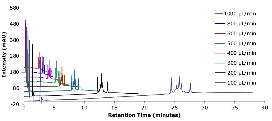
An overlayed chromatogram showing the analysis of SILu™Lite ADC Mimic on a Chromolith® WP 300 RP-18 (100 x 2 mm I.D.) column at various flow rates. The x-axis represents retention time in minutes, ranging from 0 to 40, and the y-axis represents intensity in milli-absorbance units (mAU), ranging from -20 to 580. The chromatograms are color-coded to indicate flow rates: red (1000 µL/min), yellow (800 µL/min), pink (600 µL/min), green (500 µL/min), cyan (400 µL/min), blue (300 µL/min), black (200 µL/min), and purple (100 µL/min). At higher flow rates, such as 1000 µL/min and 800 µL/min, the analyte peaks appear earlier with shorter retention times, while at lower flow rates, such as 200 µL/min and 100 µL/min, the peaks are delayed and broader, with longer retention times. The baseline becomes flatter and more stable as the flow rate decreases.

Figure 14.Analysis of SILu™Lite ADC Mimic on a Chromolith® WP 300 RP-18 (100 x 2 mm I.D.) column with different flow rates. Method: Temp. 60 °C, Mobile phase: A: Water 0.1 % (v/v) TFA, B: Acetonitrile 0.1 % (v/v) TFA; Gradient: 20% B to 60% B, adjustment see table.
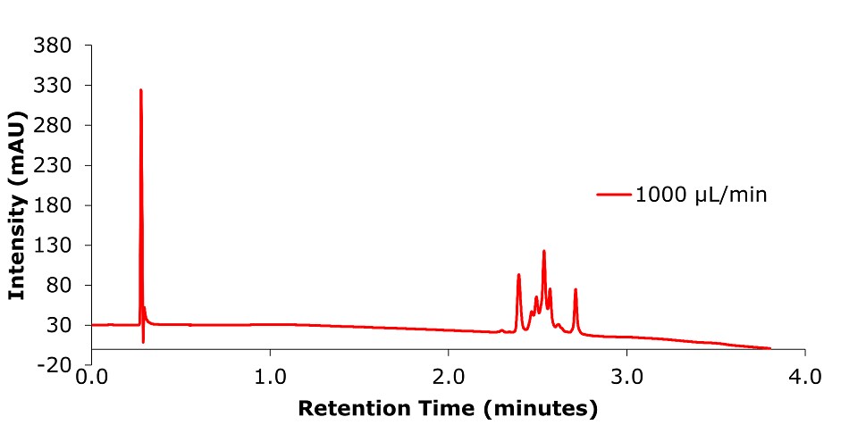
A chromatogram representing the analysis of a sample performed at a flow rate of 1000 µL/min. The x-axis indicates retention time in minutes, ranging from 0 to 4, while the y-axis represents intensity in milli-absorbance units (mAU), ranging from -20 to 380. The chromatogram, displayed as a red line, features a tall, sharp peak at the very beginning of the run near 0.3 minutes, likely representing unretained components or injection artifacts. This is followed by a relatively flat baseline, interrupted by a cluster of well-defined peaks between 2.4 and 2.7 minutes, indicating the separation of sample components.

Figure 15.Analysis of SILu™Lite ADC Mimic on a Chromolith® WP 300 RP-18 (100 x 2 mm I.D.) column at 1 mL/min.

Conclusion

Within this work, it could be shown that intact SILu™Lite SigmaMAb Universal Antibody Standard human and SigmaMAb Antibody-Drug Conjugate (ADC) Mimic could be analyzed on silica monolithic HPLC columns under various conditions.

A significant increase in peak area with temperature change from 60°C to 80°C was observed as well as with the addition of solvent additives (5% 1-Propanol or 5% 1-Butanol). The combination of 60°C with solvent additive leads to the same result as increasing the temperature to 80°C, which can be beneficial as high temperature can lead to high-temperature artifact formation. Elevating the temperature to 90°C did not show a further improvement; also, there was no significant difference between pump-mixed and pre-mixed mobile phases. Additionally, it was found that the effect of adding 1-Butanol compared to 1-Propanol leads to a different retention behavior. 1-Butanol acts as a stronger eluting solvent, leading to shorter retention time compared to acetonitrile. 1-Propanol is known to have a high affinity for surface C18 chains, which seems to keep the C18 chains completely extended1 even under high water content environments in the mobile phase, leading to an increased retention time.

Higher flow rates lead to a tremendous time saving with only minor impact on column backpressure of the silica monolithic HPLC column, which can be beneficial, especially for biomolecules, as high pressure can affect conformational changes.2 Analyzing the data related to time, peak area, and peak width are decreasing with higher flow rate. Related to volume peak area is relatively stable, which is expected for a constant concentration, and peak width is increasing due to poor mass transfer kinetics of the biomolecule. Peak height is nearly constant, decreasing only slightly with increasing flow rate.

The described effects for peak area, peak height, and peak width for increased flow rates are related to the UV detection physical principle.3,4

Find more information on Biopharmaceutical Chracterization.

Related Products
Loading

REFERENCES

1.
Lu H, Rutan S. 1999. Solvatochromic studies of solvation effects in reversed-phase liquid chromatography with addition of 1-propanol. Analytica Chimica Acta. 388(3):345-352. https://doi.org/10.1016/s0003-2670(98)00523-6
2.
Fekete S, Veuthey J, McCalley DV, Guillarme D. 2012. The effect of pressure and mobile phase velocity on the retention properties of small analytes and large biomolecules in ultra-high pressure liquid chromatography. Journal of Chromatography A. 1270127-138. https://doi.org/10.1016/j.chroma.2012.10.056
3.
Stoll DR. 2019. Effect of Flow Rate on UV Detection in Liquid Chromatography, Volume 37, Issue 12, p.846-850. [Internet]. LCGC North America: LCGC International. Available from: https://www.chromatographyonline.com/view/effects-flow-rate-uv-detection-liquid-chromatography-0
4.
Urban PL. 2016. Clarifying Misconceptions about Mass and Concentration Sensitivity. J. Chem. Educ. 93(6):984-987. https://doi.org/10.1021/acs.jchemed.5b00986
Sign In To Continue

To continue reading please sign in or create an account.

Don't Have An Account?