Western Blot Analysis of Immunoprecipitation (IP-Western)
IP-Western analysis remains a popular technique for identifying protein-protein interactions and identifying unknown proteins in a multi-protein complex. The steps include cell lysis, formation of the antibody-antigen (immune) complex, precipitation of the immune complexes, and analysis by Western blotting.
Read more about
Related Products
Loading

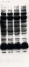
Figure 1.High background, leading to nonspecific bands on Western blot.
| Possible Cause | Remedy |
|---|---|
Non-specific binding to beads |
|
Wash buffer not stringent enough |
|
Non-specific antibody binding |
|
Inadequate washing |
|
| Possible Cause | Remedy |
|---|---|
Protein degradation, dephosphorylation, and denaturation during lysis step |
|
Lysis buffer too stringent |
|
Antibody not bound to the beads |
|
Not enough antibody-antigen binding |
|
Agarose beads accidentally removed during precipitation step |
|
Mouse IgG or chicken antibody does not bind efficiently to protein G- or protein-A conjugated beads (for indirect immunoprecipitation) |
|
Target protein remains on the beads |
|
Disruption of protein complexes (in co-immunoprecipitation experiments) |
|

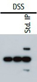
Figure 2.In a cross-linking IP western, the heavy and light chains of the primary capture antibody are excluded from the sample.
| Possible Cause | Remedy |
|---|---|
Heavy and light chains of the primary antibody are being recognized by the secondary antibody |
|
Relative Affinity | ||||||
|---|---|---|---|---|---|---|
| Antibodies | Protein A/G Mix | Protein A | Protein G | Kappa Ig Binder | Lambda Ig Binder | Kappa/Lambda Mix* |
| Rabbit IgG | SA | SA | SA | |||
| Mouse IgM | RE | SA | ||||
| Mouse IgG3 | SA | MSA | SA | |||
| Mouse IgG2b | SA | SA | SA | |||
| Mouse IgG2a | SA | SA | SA | |||
| Mouse IgG1 | MSA | MSA | MSA | |||
| Human IgM | MSA | MSA | MSA | MSA | SA | |
| Human IgE | MSA | MSA | MSA | MSA | SA | |
| Human IgD | MSA | MSA | MSA | MSA | SA | |
| Human IgA | MSA | MSA | MSA | MSA | SA | |
| Human IgG4 | SA | SA | SA | MSA | MSA | SA |
| Human IgG3 | SA | SA | MSA | MSA | SA | |
| Human IgG2 | SA | SA | SA | MSA | MSA | SA |
| Human IgG1 | SA | SA | SA | MSA | MSA | SA |
| Rat IgM | RE | RE | ||||
| Rat IgG2c | MSA | RE | MSA | |||
| Rat IgG2b | MSA | RE | MSA | |||
| Rat IgG2a | SA | RE | SA | |||
| Rat IgG1 | MSA | RE | MSA | |||
| Rat IgG | MSA | SA | SA | |||
| Fragments | Protein A/G Mix | Protein A | Protein G | Kappa Ig Binder | Lambda Ig Binder | Kappa/Lambda Mix* |
|---|---|---|---|---|---|---|
| Human l | SA | MSA | ||||
| Human k | SA | MSA | ||||
| Human Fc | MSA | MSA | MSA | |||
| Human scFv | MSA | MSA | RE | RE | RE | |
| Human F(ab')2 | MSA | MSA | MSA | MSA | MSA | SA |
| Human F(ab) | MSA | MSA | MSA | MSA | MSA | SA |
| Key code for relative affinity of protein A and G; PureProteome™ Kappa and Lambda magnetic beads for antibodies: SA = Strong Affinity; MSA = Moderate/Slight Affinity; RE = Requires Evaluation *PureProteome™ Kappa/Lambda mix is not a catalog item. Simply procure the Kappa and Lambda beads individually and mix at a 1:1 ratio. | ||||||
Sign In To Continue
To continue reading please sign in or create an account.
Don't Have An Account?