MABN1796
Anti-LRP1 Antibody, 85 kDa subunit Antibody, clone 6F8
clone 6F8, from mouse
別名:
Prolow-density lipoprotein receptor-related protein 1, A2MR, Alpha-2-macroglobulin receptor, APER, Apolipoprotein E receptor, CD19, Low-density lipoprotein receptor-related protein 1 85 kDa subunit, LRP-85
ログイン組織・契約価格を表示する
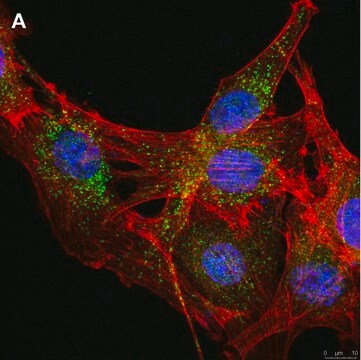
すべての画像(2)
About This Item
UNSPSCコード:
12352203
eCl@ss:
32160702
おすすめの製品
由来生物
mouse
品質水準
抗体製品の状態
purified immunoglobulin
抗体製品タイプ
primary antibodies
クローン
6F8, monoclonal
化学種の反応性
human, mouse
テクニック
ELISA: suitable
western blot: suitable
アイソタイプ
IgG1κ
NCBIアクセッション番号
輸送温度
ambient
ターゲットの翻訳後修飾
unmodified
遺伝子情報
human ... LRP1(4035)
詳細
Prolow-density lipoprotein receptor-related protein 1 (UniProt Q07954; also known as A2MR, Alpha-2-macroglobulin receptor, APER, Apolipoprotein E receptor, CD19. LRP-1) is encoded by the LRP1 (also known as A2MR, APR) gene (Gene ID 4035) in human. LRP-1 is single pass type I membrane protein that is expressed in most tissues, but is abundant in liver, brain, and lung. LRP1 is synthesized with a signal peptide sequence (a.a. 1-19) and is processed in trans-Golgi network by furin to generate a 515 kDa alpha subunit and an 85 kDa beta subunit. The alpha and beta subunits are non-covalently linked during LRP1 transport to the cell membrane. LRP1 recognizes and mediates the endocytosis of more than 40 different ligands, including apolipoprotein E (ApoE), APP and amyloid beta. It is the primary receptor mediating transport of amyloid beta peptides across the blood-brain barrier into circulation, thereby clearing them from the brain. LRP1 is required for early embryonic development and is involved in cellular lipid homeostasis and plasma clearance of chylomicron remnants and activated LRPAP1 (alpha 2-macroglobulin). Excessive copper accumulation in the brain has been linked with reduced LRP1 mediated clearance of amyloid beta peptides across the blood brain barrier, which may contribute to complications of Alzheimer′s disease.
特異性
Clone 6F8 (A.k.a. 6AF8) epitope lies within LRP1 C-terminal end sequence present in the membrane-bound beta-fragment (85 kDa subunit), but not the extracellular alpha-fragment (515 kDa subunit). Target region is 100% conserved between human and murine species, but missing in human LRP1 spliced isoform 2.
免疫原
Synthetic peptide corresponding to the C-terminal end sequence of human/mouse LRP1.
アプリケーション
Research Category
ニューロサイエンス
ニューロサイエンス
Anti-LRP1, 85 kDa subunit, clone 6F8, Cat. No. MABN1796, is a highly specific mouse monoclonal antibody that targets LRP1 85 kDa subunit and has been tested in ELISA and Western Blotting.
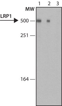
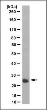
Western Blotting Analysis: 0.5 µg/mL from a representative lot detected LRP1 85 kDa subunit in 10 µg of mouse hippocampus tissue lysate.
ELISA Analysis: A representative lot was employed as the capture antibody for the detection of LRP1 in different human brain regions by sandwich ELISA. a strong positive correlation between LRP1 and PSD95 regional distribution was observed (Shinohara, M., et al. (2013). Acta Neuropathol. 125(4):535-547).
Western Blotting Analysis: A representative lot detected siRNA-mediated LRP1 knockdown in human brain vascular pericytes (Casey, C.S., et al. 2015. J. Biol. Chem. 290(22):14208-14217).
Note: The use of 5% skim milk as the blocking agent and 1-2 hr instead of overnight primary incubation time is recommended for Western blotting application to minimize non-specific background.
品質
Evaluated by Western Blotting in mouse brain and mouse hippocampus tissue lysate.
Western Blotting Analysis: 0.5 µg/mL of this antibody detected LRP1 85 kDa subunit in 10 µg of mouse whole brain tissue lysate.
Western Blotting Analysis: 0.5 µg/mL of this antibody detected LRP1 85 kDa subunit in 10 µg of mouse whole brain tissue lysate.
ターゲットの説明
~85 kDa observed. Target band size appears larger than the calculated molecular weight of 65.78 kDa (a.a. 3944-4544; UniProt Q07954) due to glycosylation. Uncharacterized bands may be observed in some lysate(s).
物理的形状
Protein G purified.
Format: Purified
Purified mouse IgG1κ in buffer containing 0.1 M Tris-Glycine (pH 7.4), 150 mM NaCl with 0.05% sodium azide.
保管および安定性
Stable for 1 year at 2-8°C from date of receipt.
その他情報
Concentration: Please refer to lot specific datasheet.
免責事項
Unless otherwise stated in our catalog or other company documentation accompanying the product(s), our products are intended for research use only and are not to be used for any other purpose, which includes but is not limited to, unauthorized commercial uses, in vitro diagnostic uses, ex vivo or in vivo therapeutic uses or any type of consumption or application to humans or animals.
適切な製品が見つかりませんか。
製品選択ツール.をお試しください
保管分類コード
12 - Non Combustible Liquids
WGK
WGK 1
適用法令
試験研究用途を考慮した関連法令を主に挙げております。化学物質以外については、一部の情報のみ提供しています。 製品を安全かつ合法的に使用することは、使用者の義務です。最新情報により修正される場合があります。WEBの反映には時間を要することがあるため、適宜SDSをご参照ください。
Jan Code
MABN1796:
試験成績書(COA)
製品のロット番号・バッチ番号を入力して、試験成績書(COA) を検索できます。ロット番号・バッチ番号は、製品ラベルに「Lot」または「Batch」に続いて記載されています。
Angela Jeong et al.
Acta neuropathologica communications, 9(1), 129-129 (2021-07-29)
The pathogenic mechanisms underlying the development of Alzheimer's disease (AD) remain elusive and to date there are no effective prevention or treatment for AD. Farnesyltransferase (FT) catalyzes a key posttranslational modification process called farnesylation, in which the isoprenoid farnesyl pyrophosphate
Dustin Chernick et al.
Journal of neurochemistry, 147(5), 647-662 (2018-07-22)
The apolipoprotein E (apoE) ε4 allele is the primary genetic risk factor for late-onset Alzheimer's disease (AD). ApoE in the brain is produced primarily by astrocytes; once secreted from these cells, apoE binds lipids and forms high-density lipoprotein (HDL)-like particles.
ライフサイエンス、有機合成、材料科学、クロマトグラフィー、分析など、あらゆる分野の研究に経験のあるメンバーがおります。.
製品に関するお問い合わせはこちら(テクニカルサービス)