추천 제품
생물학적 소스
guinea pig
Quality Level
항체 형태
serum
항체 생산 유형
primary antibodies
클론
polyclonal
종 반응성
human, mouse, rat
제조업체/상표
Chemicon®
기술
immunofluorescence: suitable
immunohistochemistry: suitable (paraffin)
western blot: suitable
NCBI 수납 번호
UniProt 수납 번호
배송 상태
dry ice
타겟 번역 후 변형
unmodified
유전자 정보
human ... SLC1A2(6506)
일반 설명
Glutamate transporters (GluT) function to remove L-glutamate (Glu), the primary excitatory neurotransmitter in the mammalian central nervous system (CNS), from the synaptic cleft. By clearing the synapse in this manner Glu can be recycled for later use, the proper diffusion gradient can be maintained and excitotoxicity can be prevented. There have been reports of many different members of a GluT multigene family; GLT1, GLAST, EAAC1 and EAAT2. mRNA has been identified in brain for each of these proteins.
EAAT2 transports L-glutamate and also L- and D-aspartate. It is essential for terminating the postsynaptic action of glutamate by rapidly removing released glutamate from the synaptic cleft. Acts as a symport by cotransporting sodium.
EAAT2 transports L-glutamate and also L- and D-aspartate. It is essential for terminating the postsynaptic action of glutamate by rapidly removing released glutamate from the synaptic cleft. Acts as a symport by cotransporting sodium.
특이성
Glial Glutamate Transporter GLT-1 (EAAT2). The antibody has been tested on central nervous system tissue. Preabsorption of the antiserum with the immunogen peptide (Catalog number AG391) completely abolishes the immunostaining.
면역원
Epitope: C-terminus
Synthetic peptide from the carboxy-terminus of rat GLT-1.
애플리케이션
Detect Glutamate Transporter using this Anti-Glutamate Transporter Antibody, Glial validated for use in IH(P), IF & WB.
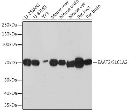
Evaluated by Western Blot on mouse brain membrane lysates.
Western Blot: 1:500 dilution of this antibody detected GLT-1 on 10 ug of mouse brain membrane lysates.
Immunohistochemistry:
1:1,000-1:4,000 dilution from a previuos lot used a DAB detection system on adult rat forebrain fixed with 4% paraformaldehyde.
Immunohistochemistry:
1:5,000-10,000 dilution from a previuos lot used a cyanine conjugated secondary antibody.
Enzymatic detection requires substantially higher primary antibody dilutions. Lightly fixed 4% PFA material is recommended.
Notes:
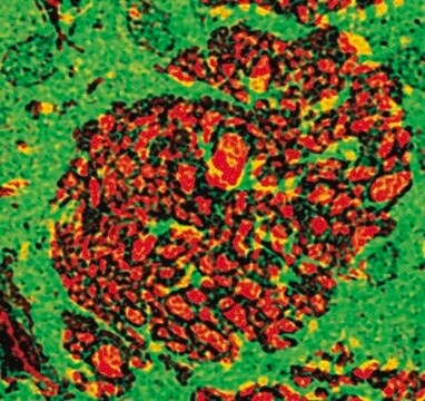
Male Sprague-Dawley rats (b.wt. 100-150g) were anesthetized with sodium pentobarbital and perfused via the ascending aorta with 50 mL of Ca2+-free Tyrode+s solution followed by a formalin-picric acid fixative (4% paraformaldehyde with 0.4% picric acid in 0.16 M phosphate buffer, pH 6.9) for 6 minutes. Tissues were rapidly dissected out, postfixed in the same fixative for 90 minutes and rinsed for at least 24 hours in 0.1 M phosphate buffer (pH 7.4) containing 10% sucrose. Sections were cut (14 um) in a cryostat and incubated at 4°C overnight with AB1783 (1:5,000-1:10,000). After rinsing in PBS sections were incubated for 60 minutes at room temperature with Cy3-conjugated secondary antibodies. After mounting in a mixture of PBS and glycerol (1:3) containing 0.1% p-phenylenediamine, sections were examined with a Nikon Microphot-SA epifluorescence microscope.
Optimal working dilutions must be determined by the end user.
Research Category
Neuroscience
Neuroscience
Research Sub Category
Ion Channels & Transporters
Ion Channels & Transporters
품질
Evaluated by Western Blot on mouse brain membrane lysates.
표적 설명
~62 kDa
물리적 형태
Guinea pig antiserum containing no preservatives.
Unpurified
저장 및 안정성
Stable for 1 year at -20ºC from date of receipt.
분석 메모
Control
Rat brain tissue
Rat brain tissue
기타 정보
Concentration: Please refer to the Certificate of Analysis for the lot-specific concentration.
법적 정보
CHEMICON is a registered trademark of Merck KGaA, Darmstadt, Germany
면책조항
Unless otherwise stated in our catalog or other company documentation accompanying the product(s), our products are intended for research use only and are not to be used for any other purpose, which includes but is not limited to, unauthorized commercial uses, in vitro diagnostic uses, ex vivo or in vivo therapeutic uses or any type of consumption or application to humans or animals.
적합한 제품을 찾을 수 없으신가요?
당사의 제품 선택기 도구.을(를) 시도해 보세요.
Storage Class Code
10 - Combustible liquids
WGK
WGK 1
시험 성적서(COA)
제품의 로트/배치 번호를 입력하여 시험 성적서(COA)을 검색하십시오. 로트 및 배치 번호는 제품 라벨에 있는 ‘로트’ 또는 ‘배치’라는 용어 뒤에서 찾을 수 있습니다.
R C Roberts et al.
Neuroscience, 277, 522-540 (2014-07-30)
The process of glutamate release, activity, and reuptake involves the astrocyte, the presynaptic and postsynaptic neurons. Glutamate is released into the synapse and may occupy and activate receptors on both neurons and astrocytes. Glutamate is rapidly removed from the synapse
Noncell-autonomous photoreceptor degeneration in a zebrafish model of choroideremia.
Krock, BL; Bilotta, J; Perkins, BD
Proceedings of the National Academy of Sciences of the USA null
Activity of D-amino acid oxidase is widespread in the human central nervous system.
Sasabe, J; Suzuki, M; Imanishi, N; Aiso, S
Frontiers in synaptic neuroscience null
Lesion-induced alterations in astrocyte glutamate transporter expression and function in the hippocampus.
Schreiner, AE; Berlinger, E; Langer, J; Kafitz, KW; Rose, CR
ISRN neurology null
Silvana Valtcheva et al.
Nature communications, 7, 13845-13845 (2016-12-21)
Astrocytes, via excitatory amino-acid transporter type-2 (EAAT2), are the major sink for released glutamate and contribute to set the strength and timing of synaptic inputs. The conditions required for the emergence of Hebbian plasticity from distributed neural activity remain elusive.
자사의 과학자팀은 생명 과학, 재료 과학, 화학 합성, 크로마토그래피, 분석 및 기타 많은 영역을 포함한 모든 과학 분야에 경험이 있습니다..
고객지원팀으로 연락바랍니다.