추천 제품
생물학적 소스
rat
Quality Level
결합
unconjugated
항체 형태
purified antibody
항체 생산 유형
primary antibodies
클론
1H2, monoclonal
분자량
calculated mol wt 56.89 kDa
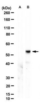
observed mol wt ~57 kDa
종 반응성
human
포장
antibody small pack of 100 μL
기술
immunofluorescence: suitable
western blot: suitable
동형
IgG2aκ
UniProt 수납 번호
배송 상태
dry ice
저장 온도
2-8°C
타겟 번역 후 변형
unmodified
일반 설명
Receptor-interacting serine/threonine-protein kinase 3 (UniProt: Q9Y572; also known as EC:2.7.11.1, RIP-like protein kinase 3, Receptor-interacting protein 3, RIP-3) is encoded by the RIPK3 (also known as RIP3) gene (Gene ID: 11035) in human. RIPK3 is a serine/threonine protein kinase that is expressed in embryo and adult spleen, liver, testis, brain, and lung. Its protein kinase domain is localized in amino acids 21 to 287 in the N-terminal half and it also has a C-terminal RIP homolytic interaction motif (RHIM; aa 450-466), which mediates interaction with the RHIM motif of RIPK1 and facilitate the formation of a necrosome. RIPK3 is shown to activate both necroptosis and apoptosis. Activated RIPK3 forms a necrosis-inducing complex and mediates phosphorylation of MLKL, promoting MLKL localization to the plasma membrane and execution of programmed necrosis that is characterized by calcium influx and plasma membrane damage. Its location is mainly cytoplasmic, however, following infection, it can also be detected in the nucleus. RIPK3 also regulates apoptosis that is dependent on RIPK1, FADD, and CASP8, and is independent of MLKL and RIPK3 kinase activity. It phosphorylates RIPK1 and then RIPK1 and RIPK3 undergo reciprocal auto- and trans-phosphorylation. In some cell types, RIPK3 is also reported to restrict viral replication by promoting cell death-independent responses. Clone 8G7 is raised against an extended kinase domain (residues 2-353) of mouse RIPK3 and can detect forms that may not be detected by antibodies directed towards the C-terminus of the full-length RIPK3. (Ref.: Samson, AL., et al. (2021). Cell Death Differ. https://doi.org/10.1038/s41418-021-00742-x; Petrie, EJ., et al. (2019). Cell Rep. 28(13); 3309-3319).
특이성
Clone 1H2 is a rat monoclonal antibody that detects human Receptor-interacting serine/threonine-protein kinase (RIP3). It targets an epitope within the extended kinase domain that includes most of the N-terminal half and a part of C-terminal half.
면역원
Recombinant fragment corresponding to 355 amino acids from the extended kinase domain of human Receptor-interacting serine/threonine-protein kinase 3 (RIPK3).
애플리케이션
Quality Control Testing
Evaluated by Western Blotting in HT-29 cell lysate.
Western Blotting Analysis: A 1:500 dilution of this antibody detected RIPK3/RIP3 in HT-29 cell lysate.
Tested Applications
Western Blotting Analysis: A representative lot detected RIPK3/RIP3 in Western Blotting applications (Samson, A.L., et. al. (2020). Nat Commun. 11(1):3151).
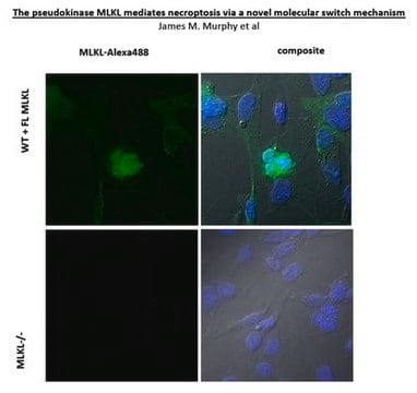
Immunofluorescence Analysis: A representative lot detected RIPK3/RIP3 in Immunofluorescence applications (Samson, A.L., et. al. (2021). Cell Death Differ. https://doi.org/10.1038/s41418-021-00742-x).
Note: Actual optimal working dilutions must be determined by end user as specimens, and experimental conditions may vary with the end user
Anti-RIPK3/RIP3, clone 1H2, Cat. No. MABC1640, is a rat monoclonal antibody that detects RIPK3/RIP3 and is tested for use in Immunofluorescence and Western Blotting.
물리적 형태
Purified rat monoclonal antibody IgG2a in buffer containing 0.1 M Tris-Glycine (pH 7.4), 150 mM NaCl with 0.05% sodium azide.
저장 및 안정성
Recommend storage at +2°C to +8°C. For long term storage antibodies can be kept at -20°C. Avoid repeated freeze-thaws.
기타 정보
Concentration: Please refer to the Certificate of Analysis for the lot-specific concentration.
면책조항
Unless otherwise stated in our catalog or other company documentation accompanying the product(s), our products are intended for research use only and are not to be used for any other purpose, which includes but is not limited to, unauthorized commercial uses, in vitro diagnostic uses, ex vivo or in vivo therapeutic uses or any type of consumption or application to humans or animals.
적합한 제품을 찾을 수 없으신가요?
당사의 제품 선택기 도구.을(를) 시도해 보세요.
Storage Class Code
12 - Non Combustible Liquids
WGK
WGK 1
Flash Point (°F)
Not applicable
Flash Point (°C)
Not applicable
시험 성적서(COA)
제품의 로트/배치 번호를 입력하여 시험 성적서(COA)을 검색하십시오. 로트 및 배치 번호는 제품 라벨에 있는 ‘로트’ 또는 ‘배치’라는 용어 뒤에서 찾을 수 있습니다.
자사의 과학자팀은 생명 과학, 재료 과학, 화학 합성, 크로마토그래피, 분석 및 기타 많은 영역을 포함한 모든 과학 분야에 경험이 있습니다..
고객지원팀으로 연락바랍니다.


