추천 제품
생물학적 소스
mouse
Quality Level
항체 형태
purified immunoglobulin
항체 생산 유형
primary antibodies
클론
RL1, monoclonal
종 반응성
human, Drosophila, Xenopus, rat
기술
affinity binding assay: suitable
electron microscopy: suitable
immunocytochemistry: suitable
immunofluorescence: suitable
immunohistochemistry: suitable
immunoprecipitation (IP): suitable
inhibition assay: suitable
western blot: suitable
동형
IgMκ
배송 상태
ambient
타겟 번역 후 변형
glycosylation
유전자 정보
human ... OGT(8473)
일반 설명
Posttranslational modification of proteins by -linked N-acetylglucosamine ( -GlcNAc) via the hydroxyl moieties on serine or threonine residues is termed O-linked -GlcNAc or simply O-GlcNAc. O-GlcNAc is one of the most abundant posttranslational modifications within the nucleocytoplasmic compartments of all animals and plants. Unlike other types of protein glycosylations, O-GlcNAc occurs exclusively within the nuclear and cytoplasmic compartments and is generally not further modified to form more elongated structures. In addition, O-GlcNAcylation is a highly dynamic and reversible process. The O-GlcNAc transferase (OGT) attaches O-GlcNAc to proteins at specific serine or threonine residues, while O-GlcNAcase catalyzes the removal/hydrolysis of O-GlcNAc from proteins. In fact, a dynamic interplay between O-GlcNAcylation and serine/threonine phosphorylation plays an important role in regulating cellular signaling. Tau and RNA polymerase II (Pol II) are two well known proteins that undergo modification by O-GlcNAcylation. In Alzheimer s diseased human brains, tau becomes extensively phosphorylated and less O-GlcNAcylated. Similarly, O-GlcNAc is removed and replaced with O-phosphate on the Poly II CTD when the elongation phase of transcription is initiated.
특이성
Both clone RL1 & clone RL2 (Cat. No. MABS157) target O-linked N-acetylglucosamine (GlcNAc) modified (GlcNAcylated) proteins. GlcNAc removal by beta-N-acetylglucosaminidase treatment or galactosylation of GlcNAc on target proteins by galactosyltransferase treatment greatly reduced the immunoreactivity of clone RL1 & RL2 toward their target proteins. The binding affinity of RL1 and RL2 toward a given GlcNAcylated protein is dependent on both O-GlcNAc and the adjacent core protein structure (Snow, C.M., et al. (1987). J. Cell Biol. 104(5):1143-11560; Holt, G.D., et al. (1987). J. Cell Biol. 104(5):1157-1164).
Target structure is not species-specific.
면역원
Nuclear pore complex-lamina fraction isolated from rat liver nuclear envelopes.
애플리케이션
Detect GlcNAcylated proteins using this mouse monoclonal Anti-O-GlcNAc, clone RL1 , Cat. No. MABS1253, validated for use in Affinity Binding, Electron Microscopy, Immunocytochemistry, Immunofluorescence, Immunohistochemistry, Immunoprecipitation, Inhibition, and Western Blotting.
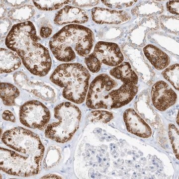
Immunohistochemistry Analysis: A 1:50 dilution from a representative lot detected O-GlcNAcylated cytoplasmic and nuclear pore proteins in rat kidney and spleen tissue sections.
Affinity Binding Assay: A representative lot of clone RL1 & clone RL2 (Cat. No. MABS157) partially competed against each other for binding immobilized 180 kDa O-GlcNAcylated nuclear envelope protein (Snow, C.M., et al. (1987). J. Cell Biol. 104(5):1143-11560).
Electron Microscopy Analysis: A representative lot immunolocalized target proteins at the cytoplasmic and/or nucleoplasmic margins of the pore complex with no specific staining of the perinuclear space of isolated rat liver nuclear envelopes (Snow, C.M., et al. (1987). J. Cell Biol. 104(5):1143-11560).
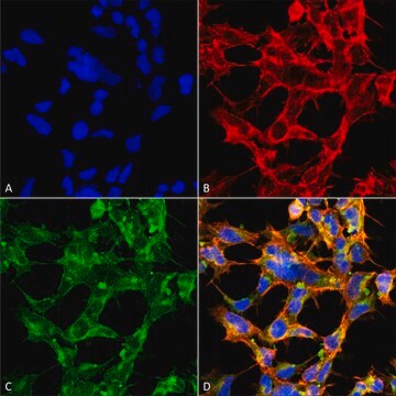
Immunocytochemistry Analysis: Representative lots stained the nuclear envelope of paraformaldehyde-fixed, Triton X-100-permeabilized HeLa cells, NRK normal rat kidney epithelial cells and isolated rat liver nuclei by fluorescent immunocytochemistry (Yang, L., et al. (1997). J. Cell Biol. 139(5):1077-1087; Byrd, D.A., et al. (1994). J. Cell Biol. 127(6 Pt 1):1515-1526; Snow, C.M., et al. (1987). J. Cell Biol. 104(5):1143-11560).
Immunofluorescence Analysis: A representative lot stained the nuclear envelope proteins in formaldehyde-fixed, Triton X-100-permeabilzied salivary glands dissected from third-instar Drosophila larvae by fluorescent immunohistochemistry (Goldberg, M., et al. (1998). Mol. Cell. Biol. 18(7):4315-4323).
Immunofluorescence Analysis: Representative lots stained the nuclear envelope of methanol-fixed xenopus ovary and rat liver cryosections by fluorescent immunohistochemistry (Featherstone, C., et al. (1988). J. Cell Biol. 107(4):1289-1297; Snow, C.M., et al. (1987). J. Cell Biol. 104(5):1143-11560).
Immunoprecipitation Analysis: A representative lot immunoprecipitated several nuclear envelope proteins in salt-washed nuclear envelope preparations from rat liver and NRK normal rat kidney epithelial cells. Galactosylation of GlcNAc on nuclear envelope proteins by galactosyltransferase treatment significantly inhibited the immunoadsorption of these nuclear pore complex glycoproteins by clone RL1 (Snow, C.M., et al. (1987). J. Cell Biol. 104(5):1143-11560; Holt, G.D., et al. (1987). J. Cell Biol. 104(5):1157-1164).
Inhibition Analysis: A representative lot, when injected in the vegetal hemisphere of xenopus oocyte cytoplasm, inhibited nucleoplasmin nuclear import in a dose-dependent manner without affecting myoglobin nuclear import or RNA export (Featherstone, C., et al. (1988). J. Cell Biol. 107(4):1289-1297).
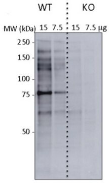
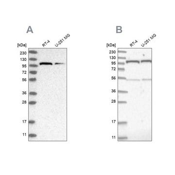
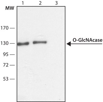
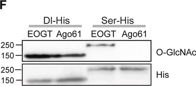
Western Blotting Analysis: Representative lots detected several nuclear envelope proteins in Xenopus oocyte nucleus extract and in salt-washed rat liver nuclear envelope preparations, including target bands of 210, 180, 145, 100, 63, 58, 54, and 45 kDa. GlcNAc removal by beta-N-acetylglucosaminidase treatment greatly reduced the detection of these nuclear pore complex glycoproteins by clone RL1 (Byrd, D.A., et al. (1994). J. Cell Biol. 127(6 Pt 1):1515-1526; Featherstone, C., et al. (1988). J. Cell Biol. 107(4):1289-1297; Snow, C.M., et al. (1987). J. Cell Biol. 104(5):1143-11560; Holt, G.D., et al. (1987). J. Cell Biol. 104(5):1157-1164).
품질
Evaluated by Immunohistochemistry in rat colon tissue.
Immunohistochemistry Analysis: A 1:50 dilution of this antibody detected O-GlcNAcylated cytoplasmic and nuclear pore proteins in rat colon tissue sections.
Immunohistochemistry Analysis: A 1:50 dilution of this antibody detected O-GlcNAcylated cytoplasmic and nuclear pore proteins in rat colon tissue sections.
표적 설명
Variable, depending on the size(s) of the O-GlcNAcylated protein(s). ~210/180/145/100/63/58/54/45 kDa reproted (Snow, C.M., et al. (1987). J. Cell Biol. 104(5):1143-11560; Holt, G.D., et al. (1987). J. Cell Biol. 104(5):1157-1164). Uncharacterized band(s) may appear in some lysates.
물리적 형태
Format: Purified
Purified mouse IgM in buffer containing PBS without preservatives.
저장 및 안정성
Stable for 1 year at -20°C from date of receipt.
Handling Recommendations: Upon receipt and prior to removing the cap, centrifuge the vial and gently mix the solution. Aliquot into microcentrifuge tubes and store at -20°C. Avoid repeated freeze
Handling Recommendations: Upon receipt and prior to removing the cap, centrifuge the vial and gently mix the solution. Aliquot into microcentrifuge tubes and store at -20°C. Avoid repeated freeze
기타 정보
Concentration: Please refer to lot specific datasheet.
적합한 제품을 찾을 수 없으신가요?
당사의 제품 선택기 도구.을(를) 시도해 보세요.
Storage Class Code
12 - Non Combustible Liquids
WGK
WGK 2
Flash Point (°F)
Not applicable
Flash Point (°C)
Not applicable
시험 성적서(COA)
제품의 로트/배치 번호를 입력하여 시험 성적서(COA)을 검색하십시오. 로트 및 배치 번호는 제품 라벨에 있는 ‘로트’ 또는 ‘배치’라는 용어 뒤에서 찾을 수 있습니다.
자사의 과학자팀은 생명 과학, 재료 과학, 화학 합성, 크로마토그래피, 분석 및 기타 많은 영역을 포함한 모든 과학 분야에 경험이 있습니다..
고객지원팀으로 연락바랍니다.