추천 제품
애플리케이션
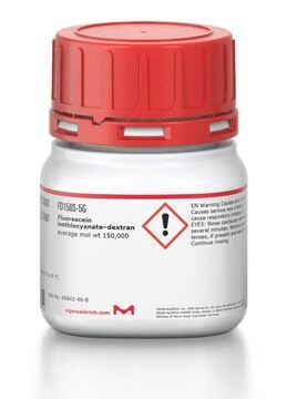
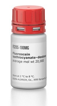
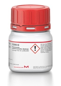
Dextran labeled with fluorescein isothiocyanate for possible use in perfusion studies in animals.
Fluorescein isothiocyanate–dextran (FITC-Dextran 150) is use as a fluorescent probe to study cell permeability. FITC-Dextran 150 is useful to study processes that affect the permeability of the blood brain barrier (BBB) and microvascular structures. FITC-Dextran 150 is used in fluorescence microlymphography.
기타 정보
Commonly utilized as a macromolecular fluorochrome in inflammatory studies
신호어
Warning
유해 및 위험 성명서
Hazard Classifications
Eye Irrit. 2 - Skin Irrit. 2 - STOT SE 3
표적 기관
Respiratory system
Storage Class Code
11 - Combustible Solids
WGK
WGK 3
Flash Point (°F)
Not applicable
Flash Point (°C)
Not applicable
개인 보호 장비
Eyeshields, Gloves, type N95 (US)
시험 성적서(COA)
제품의 로트/배치 번호를 입력하여 시험 성적서(COA)을 검색하십시오. 로트 및 배치 번호는 제품 라벨에 있는 ‘로트’ 또는 ‘배치’라는 용어 뒤에서 찾을 수 있습니다.
이미 열람한 고객
D M Gawlowski et al.
Microvascular research, 37(1), 1-15 (1989-01-01)
Fluorescein-labeled dextran MW 150K (FITC-Dx 150) is commonly utilized as a macromolecular fluorochrome in inflammatory studies. We examined the influence of FITC-Dx 150 on leukocyte activity as assessed by adherence to cheek pouch microvessels. Transcapillary exchange was evaluated as fluorochrome
Fátima Z G A Cyrino et al.
Clinical and experimental pharmacology & physiology, 31(3), 159-162 (2004-03-11)
1. The present study was designed to evaluate the effect of micronization on the protective effect of the purified flavonoid fraction (MPFF) on increases in macromolecular permeability induced by ischaemia-reperfusion in the hamster cheek pouch microcirculation. 2. Male hamsters (Mesocricetus
Haruo Aramoto et al.
American journal of physiology. Heart and circulatory physiology, 287(4), H1590-H1598 (2004-05-25)
Vascular endothelial growth factor (VEGF) induces mild vasodilation and strong increases in microvascular permeability. Using intravital microscopy and digital integrated optical intensity image analysis, we tested, in the hamster cheek pouch microcirculation, the hypothesis that differential signaling pathways in arterioles
A Bollinger et al.
Lymphology, 40(2), 52-62 (2007-09-15)
Fluorescence microlymphography (FML) is an almost atraumatic technique used to visualize the superficial skin network of initial lymphatics through the intact skin of man. Visualization was performed with an incident light fluorescence microscope following subepidermal injection of minute amounts of
Neil Y C Lin et al.
Proceedings of the National Academy of Sciences of the United States of America, 116(12), 5399-5404 (2019-03-06)
Three-dimensional renal tissues that emulate the cellular composition, geometry, and function of native kidney tissue would enable fundamental studies of filtration and reabsorption. Here, we have created 3D vascularized proximal tubule models composed of adjacent conduits that are lined with
자사의 과학자팀은 생명 과학, 재료 과학, 화학 합성, 크로마토그래피, 분석 및 기타 많은 영역을 포함한 모든 과학 분야에 경험이 있습니다..
고객지원팀으로 연락바랍니다.
