추천 제품
생물학적 소스
mouse
Quality Level
결합
unconjugated
항체 형태
purified from hybridoma cell culture
항체 생산 유형
primary antibodies
클론
CY-96, monoclonal
양식
buffered aqueous solution
농도
~1.5 mg/mL
기술
direct ELISA: suitable
dot blot: 1-2 μg/mL using cell protein extracts labeded with Cy3 or Cy5
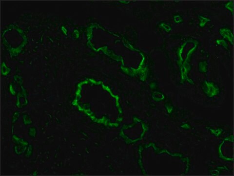
immunocytochemistry: suitable
immunoprecipitation (IP): suitable
microarray: suitable
동형
IgG2a
배송 상태
dry ice
저장 온도
−20°C
타겟 번역 후 변형
unmodified
유사한 제품을 찾으십니까? 방문 제품 비교 안내
일반 설명
Anti-Cy3/Cy5 antibody, Mouse monoclonal, (mouse IgG2a isotype) is derived from the hybridoma CY-96 produced by the fusion of mouse myeloma cells (NS1 cells) and splenocytes from Balb/c mice immunized with a mixture of proteins labeled with Cy3 or Cy5.
특이성
The antibody recognizes Cy3 and Cy5 conjugated to proteins.
면역원
mixture of proteins labeled with Cy3/Cy5.
애플리케이션
Anti-Cy3/Cy5 antibody, Mouse monoclonal has been used in:
- immunofluorescence
- western blot
- dot blot
- enzyme linked immunosorbent assay (ELISA)
- immunoprecipitation
- immunocytochemistry
- protein microarrays
- In in situ hybridization
Applications in which this antibody has been used successfully, and the associated peer-reviewed papers, are given below.
Immunofluorescence (1 paper)
Immunofluorescence (1 paper)
생화학적/생리학적 작용
Cy 3 and Cy5 are the most popular cyanine dyes, used combined for two color detection. Cy3 dyes are fluorescent orange while Cy5 is fluorescent in the red region. Cyanine belonging to polymethine group. CyDyes are a family of fluorophores that can be used for labeling proteins, peptides, DNA, RNA, and other biomolecules. These dyes are small, pH insensitive, soluble in aqueous solution and are tolerant to DMSO. They are more photostable than fluorescein, have high molar extinction coefficients and favorable quantum yields. Mainly Cy3 and Cy5 are used in many different biological assays such as DNA microarrays, protein microarrays, two-dimensional protein analysis (2D gels), fluorescence resonance energy transfer (FRET), and immunocytochemistry.
물리적 형태
Solution in 0.01 M phosphate buffered saline, pH 7.4, containing 15 mM sodium azide.
면책조항
Unless otherwise stated in our catalog or other company documentation accompanying the product(s), our products are intended for research use only and are not to be used for any other purpose, which includes but is not limited to, unauthorized commercial uses, in vitro diagnostic uses, ex vivo or in vivo therapeutic uses or any type of consumption or application to humans or animals.
적합한 제품을 찾을 수 없으신가요?
당사의 제품 선택기 도구.을(를) 시도해 보세요.
Storage Class Code
10 - Combustible liquids
WGK
WGK 3
Flash Point (°F)
Not applicable
Flash Point (°C)
Not applicable
개인 보호 장비
Eyeshields, Gloves, multi-purpose combination respirator cartridge (US)
가장 최신 버전 중 하나를 선택하세요:
시험 성적서(COA)
Lot/Batch Number
A K Kenworthy
Methods (San Diego, Calif.), 24(3), 289-296 (2001-06-14)
Fluorescence resonance energy transfer (FRET) detects the proximity of fluorescently labeled molecules over distances >100 A. When performed in a fluorescence microscope, FRET can be used to map protein-protein interactions in vivo. We here describe a FRET microscopy method that
Transplantation of ovarian granulosa-like cells derived from human induced pluripotent stem cells for the treatment of murine premature ovarian failure
Liu T. et al.
Molecular Medicine Reports, 13(6), 5053-5058 (2016)
An Improved Method for Cell Type-Selective Glycomic Analysis of Tissue Sections Assisted by Fluorescence Laser Microdissection
Nagai-Okatani C, et al.
International Journal of Molecular Sciences, 20(3), 700-700 (2019)
G Enders
Acta neurochirurgica. Supplement, 89, 9-13 (2004-09-01)
Microarray analysis has been emerged as a tool to characterize the overall reaction of cells in culture or tissue to different stimuli e.g. stressful events by analysing bulk RNA present at a particular time point. It has supplemented or even
Chiaki Nagai-Okatani et al.
International journal of molecular sciences, 20(3) (2019-02-10)
Lectin microarray (LMA) is a highly sensitive technology used to obtain the global glycomic profiles of endogenous glycoproteins in biological samples including formalin-fixed paraffin-embedded tissue sections. Here, we describe an effective method for cell type-selective glycomic profiling of tissue fragments
자사의 과학자팀은 생명 과학, 재료 과학, 화학 합성, 크로마토그래피, 분석 및 기타 많은 영역을 포함한 모든 과학 분야에 경험이 있습니다..
고객지원팀으로 연락바랍니다.


