
추천 제품


일반 설명
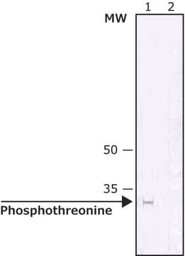
As determined by ELISA and dot blot, the antibody reacts specifically with phosphorylated threonine, both as free amino acid or conjugated to carriers such as BSA or KLH. No cross-reactivity is observed with non-phosphorylated threonine, phosphoserine, phosphotyrosine, AMP or ATP. This antibody has been used in immunoblotting for the localization of some phosphothreonine-containing proteins. Certain proteins known to contain phosphorylated threonine may not be recognized by this antibody due to steric hindrance of the recognition site.
Monoclonal Anti-Phosphothreonine (mouse IgG2b isotype) is derived from the hybridoma produced by the fusion of mouse myeloma cells and splenocytes from an immunized mouse.
면역원
phosphothreonine conjugated to keyhole limpet hemocyanin (KLH).
애플리케이션
Monoclonal Anti-Phosphothreonine antibody produced in mouse has been used in:immunoblotting, enzyme linked immuno sorbent assay (ELISA) ,dot blot.
Monoclonal and polyclonal antibodies directed against phosphorylated residues may be useful as analytical and preparative tools, by enabling the identification, quantification and immunoaffinity isolation of phosphorylated cellular proteins. Antibodies can be employed to monitor alterations in phosphorylation of specific proteins as they occur in intact organs or even whole animals.
Mouse monoclonal clone PTR-8 anti-phosphothreonine antibody may be used for the localization of phosphorylated threonine using various immunochemical assays such as ELISA, dot blot, and immunoblotting. Due to steric hindrance of the recognition site, this antibody may not recognize certain proteins known to contain phosphorylated threonine.
Mouse monoclonal clone PTR-8 anti-phosphothreonine antibody may be used for the localization of phosphorylated threonine using various immunochemical assays such as ELISA, dot blot, and immunoblotting. Due to steric hindrance of the recognition site, this antibody may not recognize certain proteins known to contain phosphorylated threonine.
Mouse monoclonal clone PTR-8 anti-Phosphothreonine antibody reacts with phosphorylated threonine both as a free amino acid or when conjugated to carriers such as BSA or KLH, using ELISA and dot blot. It does not react with nonphosphorylated threonine, phosphorylated tyrosine or serine, AMP or ATP.
생화학적/생리학적 작용
Protein phosphorylation and dephosphorylation are basic mechanisms for the modification of protein function in eukaryotic cells. Phosphorylation is a rare post-translational event in normal tissue. However, the abundance of phosphorylated cellular proteins increases tenfold following various activation processes, which are mediated through phosphotyrosine, phosphoserine or phosphothreonine (p-Tyr/p-Ser/p-Thr). Many different mitogenic systems, such as the EGF, PDGF and insulin receptor systems, contain Tyr/Ser/Thr kinase domains that autophosphorylate specific Tyr/Ser/Thr residues upon binding of their ligands. T cell antigen receptor complex or receptors for some hemopoietic growth factors may stimulate associated kinases, and cells transformed by viral oncogenes contain elevated levels of phosphorylated Tyr/Ser/Thr. An understanding of transformation by oncogenes and mitogenic processes of growth factors depends on the identification of their substrate and a subsequent determination of how phosphorylation affects the properties of these proteins.
면책조항
Unless otherwise stated in our catalog or other company documentation accompanying the product(s), our products are intended for research use only and are not to be used for any other purpose, which includes but is not limited to, unauthorized commercial uses, in vitro diagnostic uses, ex vivo or in vivo therapeutic uses or any type of consumption or application to humans or animals.
적합한 제품을 찾을 수 없으신가요?
당사의 제품 선택기 도구.을(를) 시도해 보세요.
Storage Class Code
10 - Combustible liquids
WGK
WGK 3
Flash Point (°F)
Not applicable
Flash Point (°C)
Not applicable
이미 열람한 고객
Quantification of the Dynamic Phosphorylation Process Of ERK Using Stable Isotope Dilution Selective Reaction Monitoring Mass Spectrometry
Lee N, et al.
Proteomics, 1900086-1900086 (2019)
Clémence Rougeaux et al.
Cellular microbiology, 10(3), 632-654 (2007-11-06)
Human decay accelerating factor (hDAF, CD55) and members of the carcinoembryonic-antigen-related cell-adhesion molecules (hCEACAMs) family are recognized as receptors by Gram-negative, diffusely adhering Escherichia coli (DAEC) strains expressing Afa/Dr adhesins. We report here that hCEACAM1-4L has a key function in
Sijo V Chemmannur et al.
International journal of nanomedicine, 11, 2039-2051 (2016-06-09)
Owing to the suppression of immune responses and associated side effects, steroid based treatments for inflammatory encephalitis disease can be detrimental. Here, we demonstrate a novel carbon nanosphere (CNP) based treatment regime for encephalomyelitis in mice by exploiting the functional
S L Gaffen et al.
Proceedings of the National Academy of Sciences of the United States of America, 92(16), 7192-7196 (1995-08-01)
To explore the possible involvement of STAT factors ("signal transducers and activators of transcription") in the interleukin 2 receptor (IL-2R) signaling cascade, murine HT-2 cells expressing chimeric receptors composed of the extracellular domain of the erythropoietin receptor fused to the
Takeru Zama et al.
The Journal of biological chemistry, 277(26), 23909-23918 (2002-04-18)
Mitogen-activated protein kinases (MAPKs) are activated in response to various extracellular stimuli, and their activities are regulated by upstream activating kinases and protein phosphatases such as MAPK phosphatases (MKPs). We report the identification and characterization of a novel MKP termed
자사의 과학자팀은 생명 과학, 재료 과학, 화학 합성, 크로마토그래피, 분석 및 기타 많은 영역을 포함한 모든 과학 분야에 경험이 있습니다..
고객지원팀으로 연락바랍니다.