05-321
Anti-Phosphotyrosine Antibody, clone 4G10®
clone 4G10®, Upstate®, from mouse
Synonym(s):
Anti-phosphotyrosine clone 4G10
About This Item
Recommended Products
biological source
mouse
Quality Level
antibody form
purified antibody
antibody product type
primary antibodies
clone
4G10®, monoclonal
species reactivity (predicted by homology)
all
manufacturer/tradename
Upstate®
technique(s)
immunocytochemistry: suitable
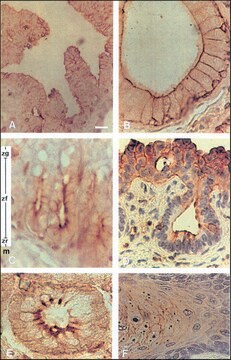
immunohistochemistry: suitable
immunoprecipitation (IP): suitable
western blot: suitable
isotype
IgG2bκ
shipped in
wet ice
General description
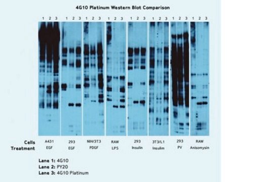
The advent of anti-phosphotyrosine antibodies is one of significant events in signal transduction research. Before the availability of anti-phosphotyrosine antibodies, tyrosyl phospyhorylation of proteins and enzymes was investigated through hazardous and time-consuming radioactive experiments. Anti-phosphotyrosine antibodies are commonly used in western blots after the targeted proteins have been immunoprecipitated to measure the tyrosyl phosphorylation of the proteins. Anti-phosphotyrosine antibodies are also directly used on cell lysate to examine the overall change of tyrosine phosphorylation level in reponse to various treatments.
Specificity
Immunogen
Application
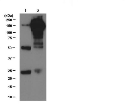
2-4 μg of a previous lot immunoprecipitated quantitatively the phosphotyrosine containing proteins in the lysate of a confluent culture (10 cm dish) of cells expressing an activated tyrosine kinase. To preserve phosphotyrosine, add: 0.2 mM sodium orthovanadate to the lysis buffer.
Signaling
General Post-translation Modification
Quality
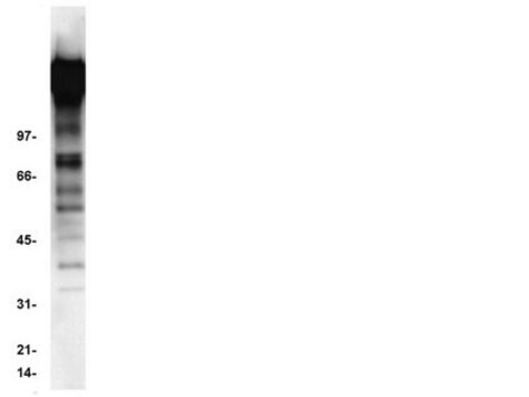
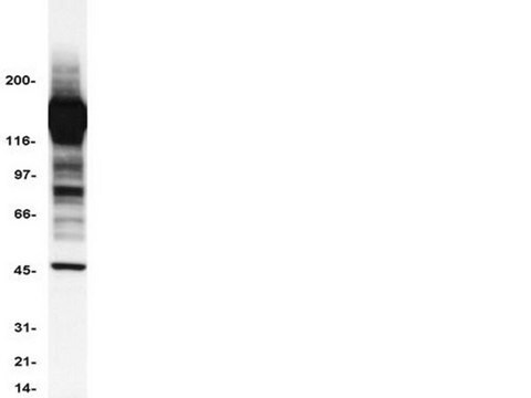
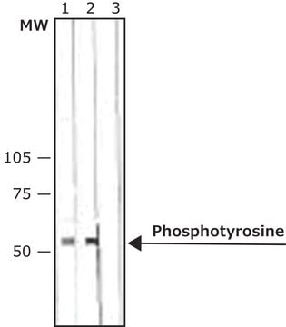
Western Blot Analysis:
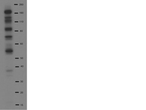
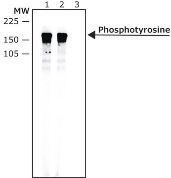
0.5-2 μg/mL of this lot detected tyrosine-phosphorylated proteins in a modified RIPA lysate from EGF-treated human A431 carcinoma cells (Cohen, B., 1990; , Druker, B. J., 1989; Kanakura, Y., 1991).
Target description
Linkage
Physical form
IgG2bκ mouse monocolonal antibody produced in vitro by mouse-mouse hybridoma 4G10 (FOX-NY [NS-1 derivative] myeloma x spleen cells).
Storage and Stability
NOTE: DO NOT FREEZE.
For maximum recovery of the product, centrifuge the original vial prior to removing the cap. If the product has accidentally been frozen and thawed, spin it at 13,000 x g for 10 minutes at 2-8ºC. Save the supernatant for application.
Analysis Note
Positive Antigen Control: Catalog #12-302, EGF-stimulated A431 cell lysate. Add 2.5µL of 2-mercaptoethanol/100µL of lysate and boil for 5 minutes to reduce the preparation. Load 20µg of reduced lysate per lane for minigels.
Other Notes
Legal Information
Disclaimer
Not finding the right product?
Try our Product Selector Tool.
recommended
Storage Class
12 - Non Combustible Liquids
wgk_germany
WGK 1
flash_point_f
Not applicable
flash_point_c
Not applicable
Certificates of Analysis (COA)
Search for Certificates of Analysis (COA) by entering the products Lot/Batch Number. Lot and Batch Numbers can be found on a product’s label following the words ‘Lot’ or ‘Batch’.
Already Own This Product?
Find documentation for the products that you have recently purchased in the Document Library.
Customers Also Viewed
Articles
Immunofluorescence uses antibody-conjugated fluorescent molecules for protein localization, modification confirmation, and protein complex visualization.
Protocols
Tips and troubleshooting for FFPE and frozen tissue immunohistochemistry (IHC) protocols using both brightfield analysis of chromogenic detection and fluorescent microscopy.
Our team of scientists has experience in all areas of research including Life Science, Material Science, Chemical Synthesis, Chromatography, Analytical and many others.
Contact Technical Service