MAB10754
Anti-Caspase-8 (active form p18 subunit) Antibody, clone 2B12.1
clone 2B12.1, from mouse
Sign Into View Organizational & Contract Pricing
All Photos(2)
caspase 8, apoptosis-related cysteine peptidase, caspase-8, caspase 8, apoptosis-related cysteine protease, MACH-beta-1/2/3/4 protein, Apoptotic protease Mch-5, Apoptotic cysteine protease, MACH-alpha-1/2/3 protein, FADD-homologous ICE/ced-3-like proteas
Recommended Products
biological source
mouse
Quality Level
antibody form
purified immunoglobulin
antibody product type
primary antibodies
clone
2B12.1, monoclonal
species reactivity
human
technique(s)
immunocytochemistry: suitable
western blot: suitable
isotype
IgMκ
NCBI accession no.
UniProt accession no.
shipped in
wet ice
target post-translational modification
unmodified
Gene Information
human ... CASP8(841)
General description
Caspase-8 (MACH/FLICE/Mch5) is a member of a family of cysteine-requiring aspartate proteases that participate in the intracellular signaling cascade leading to apoptosis, or programmed cell death. Caspase-8 is recruited to death-inducing signaling complex (DISC), CD95 (Fas/APO-1) and tumor necrosis factor receptor 1 (TNFR1) and is subsequently activated by proteolytic cleavage into two residues, p10 and p18. Active caspase-8 or the p18 subunit, is the first in the caspase signaling cascade, and it signals downstream caspases to eventually induce apoptosis.
Specificity
This antibody recognizes the active form (p18 subunit) of Caspase-8.
Immunogen
KLH-conjugated linear peptide corresponding to the active form of human Caspase-8.
Application
Detect Caspase-8 (active form p18 subunit) using this Anti-Caspase-8 (active form p18 subunit) Antibody, clone 2B12.1 validated for use in WB & IC.
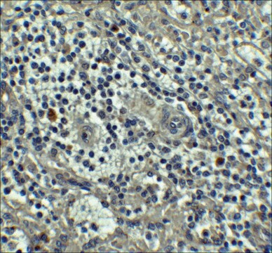
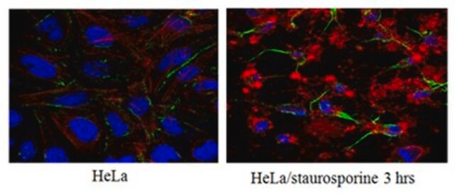
Immunocytochemistry Analysis: A 1:500 dilution from a previous lot detected active Caspase-8 (p18 subunit) in etoposide-treated HeLa cells.
Research Category
Apoptosis & Cancer
Apoptosis & Cancer
Research Sub Category
Caspases
Apoptosis - Additional
Caspases
Apoptosis - Additional
Quality
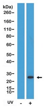
Evaluated by Western Blot in camptothecin-treated and untreated Jurkat lysates.
Western Blot Analysis: 2 µg/mL of this antibody detected the active form of Caspase-8 (p18 subunit) in 10 µg of camptothecin-treated Jurkat lysates. Untreated jurkat lysate exhibited no detection of active Caspase-8.
Target description
~18 kDa observed
Linkage
Replaces: 04-573; 04-574
Physical form
Format: Purified
Purified mouse monoclonal IgMκ in buffer containing PBS with 0.05% sodium azide.
Storage and Stability
Stable for 1 year at 2-8°C from date of receipt.
Analysis Note
Control
Camptothecin-treated and untreated Jurkat lysates
Other Notes
Concentration: Please refer to the Certificate of Analysis for the lot-specific concentration.
Disclaimer
Unless otherwise stated in our catalog or other company documentation accompanying the product(s), our products are intended for research use only and are not to be used for any other purpose, which includes but is not limited to, unauthorized commercial uses, in vitro diagnostic uses, ex vivo or in vivo therapeutic uses or any type of consumption or application to humans or animals.
recommended
Product No.
Description
Pricing
wgk_germany
WGK 2
flash_point_f
Not applicable
flash_point_c
Not applicable
Certificates of Analysis (COA)
Search for Certificates of Analysis (COA) by entering the products Lot/Batch Number. Lot and Batch Numbers can be found on a product’s label following the words ‘Lot’ or ‘Batch’.
Already Own This Product?
Find documentation for the products that you have recently purchased in the Document Library.
Nature communications, 13(1), 166-166 (2022-01-12)
Muscle cell death in polymyositis is induced by CD8+ cytotoxic T lymphocytes. We hypothesized that the injured muscle fibers release pro-inflammatory molecules, which would further accelerate CD8+ cytotoxic T lymphocytes-induced muscle injury, and inhibition of the cell death of muscle
Neurobiology of disease, 67, 49-56 (2014-03-25)
Epigenetic mechanisms are fundamental for shaping the activity of the central nervous system (CNS). Methyl-CpG binding protein 2 (MECP2) acts as a bridge between methylated DNA and transcriptional effectors responsible for differentiation programs in neurons. The importance of MECP2 dosage
Our team of scientists has experience in all areas of research including Life Science, Material Science, Chemical Synthesis, Chromatography, Analytical and many others.
Contact Technical Service