
MAB3560
Anti-8-Oxoguanine Antibody, clone 483.15
ascites fluid, clone 483.15, Chemicon®
Synonym(s):
8-oxoG
Sign Into View Organizational & Contract Pricing
Select a Size
All Photos(1)
Select a Size
Change View
About This Item
UNSPSC Code:
12352203
eCl@ss:
32160702
NACRES:
NA.41
Recommended Products


biological source
mouse
Quality Level
antibody form
ascites fluid
antibody product type
primary antibodies
clone
483.15, monoclonal
species reactivity
monkey, human, primate
species reactivity (predicted by homology)
mouse, rat
manufacturer/tradename
Chemicon®
technique(s)
ChIP: suitable
ELISA: suitable
immunocytochemistry: suitable
isotype
IgM
General description
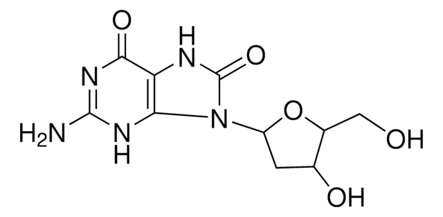
8-Oxoguanine is a mutagenic oxidative damage product of guanine. Oxidatively damaged base 8-oxoguanine is generated in the DNA of all living organisms due to the presence of reactive oxygen species in cells. DNA damage threatens the health of the genetic information stored in every DNA molecule; however enzymes exist in cells to protect against the mutagenic effect of this lesion. 8-oxoguanine is one of the most abundant and well-characterized DNA lesions generated by oxidative stress. It has been estimated that ~180 guanines are oxidized to 8-oxoG per mammalian cell per day. 8-oxoG is a miscoding lesion that can cause G:C to T:A or T:A to G:C transversion mutations. This lesion accumulates in DNA with age and it has been loosly linked to several cancers and diseases, such as Alzheimer′s and Parkinson′s.
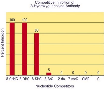
Specificity
8-oxoguanine. By competitive ELISA the monoclonal showed no reactivity with dGMP, dAMP, dCMP or TMP in micromolar concentrations. Competition was only observed when the concentrations were increased to the millimolar range.
Immunogen
8-oxoguanine adsorbed on to alumina
Application
Anti-8-Oxoguanine Antibody, clone 483.15 detects level of 8-Oxoguanine & has been published & validated for use in ELISA & IC.
Immunocytochemistry:
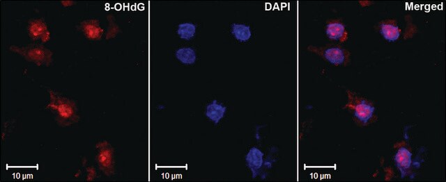
On HeLa and Cos7 cells fixed with paraformaldehyde. 8-oxoguanine has been localized to the nucleus in nutrient-deprived cells.
ELISA:
Cell grown in slides were extracted twice for 30 sec with cold 560nM NaCl;0.1% (v/v) Triton X-100;0.02% (w/v) SDS;10mM phosphate buffer pH 7.4 and fixed with freshly prepared 4% PFA for 5-20 minutes at room temperature. {Conlon, KA et al (2000) J. Histotechnology 23(1):37-44}. Typical staining shows that nuclei from extracted cells have define periphery and areas of condensed chromatin. Clone 483.15 staining showed specific but faint nuclear fluorescence staining in cells incubated in supplemented DMEM, but in cells incubated in nutrient-free defined solt solution (NFDSS) {1.8mM calcium chloride, 110mM NaCl, 44mM sodium biocarbonate, pH 7.5} showed strong nuclear fluorescence staining that appeared punctate and gernerally distributed. Fluorescence staining disappears to background levels in cells incubated in nutrient medias even after initial NFDSS treatments {Conlon et al}.
A previous lot of this antibody was used in an ELISA assay.
Optimal working dilutions must be determined by end user.
Quality
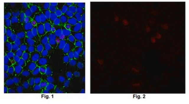
Routinely evaluated by immunocytochemistry on NIH/3T3 cells..
Immunocytochemistry:
Confocal fluorescent analysis of NIH/3T3 cells using anti-8-oxoguanine mouse monoclonal antibody.
Immunocytochemistry:
Confocal fluorescent analysis of NIH/3T3 cells using anti-8-oxoguanine mouse monoclonal antibody.
Physical form
Unpurified mouse monoclonal IgM liquid in buffer containing no preservative.
Storage and Stability
Stable for 6 months at -20ºC in undiluted aliquots from date of receipt.
Handling Recommendations: Upon receipt, and prior to removing the cap, centrifuge the vial and gently mix the solution. Aliquot into microcentrifuge tubes and store at -20°C. Avoid repeated freeze/thaw cycles, which may damage IgM and affect product performance.
Handling Recommendations: Upon receipt, and prior to removing the cap, centrifuge the vial and gently mix the solution. Aliquot into microcentrifuge tubes and store at -20°C. Avoid repeated freeze/thaw cycles, which may damage IgM and affect product performance.
Analysis Note
Control
Nutrient starved HeLa or Cos7 cells
Nutrient starved HeLa or Cos7 cells
Other Notes
Concentration: Please refer to the Certificate of Analysis for the lot-specific concentration.
Legal Information
CHEMICON is a registered trademark of Merck KGaA, Darmstadt, Germany
Not finding the right product?
Try our Product Selector Tool.
Storage Class
10 - Combustible liquids
wgk_germany
nwg
flash_point_f
Not applicable
flash_point_c
Not applicable
Certificates of Analysis (COA)
Search for Certificates of Analysis (COA) by entering the products Lot/Batch Number. Lot and Batch Numbers can be found on a product’s label following the words ‘Lot’ or ‘Batch’.
Already Own This Product?
Find documentation for the products that you have recently purchased in the Document Library.
Vascular effects of a low-carbohydrate high-protein diet.
Foo, SY; Heller, ER; Wykrzykowska, J; Sullivan, CJ; Manning-Tobin, JJ; Moore et al.
Proceedings of the National Academy of Sciences of the USA null
Robert Lowe et al.
Genome biology, 16, 194-194 (2015-09-19)
Cellular senescence is a stable arrest of proliferation and is considered a key component of processes associated with carcinogenesis and other ageing-related phenotypes. Here, we perform methylome analysis of actively dividing and deeply senescent normal human epithelial cells. We identify
Sameera Nallanthighal et al.
Nanotoxicology, 11(8), 996-1011 (2017-10-20)
Due to extensive use in consumer goods, it is important to understand the genotoxicity of silver nanoparticles (AgNPs) and identify susceptible populations. 8-Oxoguanine DNA glycosylase 1 (OGG1) excises 8-oxo-7,8-dihydro-2-deoxyguanine (8-oxoG), a pro-mutagenic lesion induced by oxidative stress. To understand whether
Ilir Mehmeti et al.
Biochimica et biophysica acta, 1813(10), 1827-1835 (2011-07-26)
Pro-inflammatory cytokine-mediated beta cell apoptosis is activated through multiple signaling pathways involving mitochondria and endoplasmic reticulum. Activation of organelle-specific caspases has been implicated in the progression and execution of cell death. This study was therefore performed to elucidate the effects
Christos Polytarchou et al.
Molecular and cellular biology, 28(24), 7451-7464 (2008-10-08)
The histone H3 demethylase Ndy1/KDM2B protects cells from replicative senescence. Changes in the metabolism of reactive oxygen species (ROS) are important for establishing senescence, suggesting that Ndy1 may play a role in redox regulation. Here we show that Ndy1 protects
Our team of scientists has experience in all areas of research including Life Science, Material Science, Chemical Synthesis, Chromatography, Analytical and many others.
Contact Technical Service