Skip to Content
MilliporeSigma
All Photos(1)

Key Documents

MABE1104M

Sigma-Aldrich

Anti-RAG-2 Antibody, clone 39

culture supernatant, clone 39, from rabbit

Synonym(s):

V(D)J recombination-activating protein 2, RAG-2

Sign Into View Organizational & Contract Pricing


About This Item

UNSPSC Code:
12352203
eCl@ss:
32160702
NACRES:
NA.41

biological source

rabbit

Quality Level

antibody form

culture supernatant

antibody product type

primary antibodies

clone

39, monoclonal

species reactivity

mouse

technique(s)

ChIP: suitable
immunofluorescence: suitable
immunoprecipitation (IP): suitable
western blot: suitable

NCBI accession no.

UniProt accession no.

shipped in

ambient

target post-translational modification

unmodified

Gene Information

mouse ... Rag2(19374)

General description

V(D)J recombination-activating protein 2 (UniProt: P21784; also known as RAG-2) is encoded by the Rag2 (also known as Rag-2) gene (Gene ID: 19374) in murine species. RAG-2 is a nuclear protein that is a core component of the multiprotein RAG complex. It is predominantly found in maturing lymphoid cells. RAG complex mediates the DNA cleavage phase during V(D)J recombination. During this phase V(D)J recombination assembles a diverse repertoire of immunoglobulin and T-cell receptor genes in developing B and T-lymphocytes through rearrangement of different V (variable), in some cases D (diversity), and J (joining) gene segments. DNA cleavage by the RAG complex is reported to occur by first introducing a nick in the top strand immediately upstream of the heptamer, generating a 3′-hydroxyl group that can attack the phosphodiester bond on the opposite strand in a direct trans-esterification reaction, thereby creating 4 DNA ends: 2 hairpin coding ends and 2 blunt, 5′-phosphorylated ends. The chromatin structure is shown to play a critical role in the V(D)J recombination reactions and the presence of histone H3 trimethylated at Lys4 (H3K4me3) stimulates both the nicking and hairpinning steps. RAG-2 contains an atypical PHD-type zinc finger (a 416-484) that recognizes and binds histone H3 trimethylated on Lys4 (H3K4me3). The introduction of DNA breaks by the RAG complex on one immunoglobulin allele induces ATM-dependent repositioning of the other allele to pericentromeric heterochromatin, preventing accessibility to the RAG complex and recombination of the second allele. Although RAG-2 is not the catalytic component, it is required for all known catalytic activities mediated by RAG1. Mice with defective Rag2 gene fail to produce mature B or T-lymphocytes.

Specificity

Clone 39 is a rabbit monoclonal antibody that detects RAG-2 protein in murine species.

Immunogen

GST-tagged recombinantn fragment corresponind to the first 490 amino acids of murine V(D)J recombination-activating protein 2 (RAG-2).

Application

Anti-RAG-2, clone 39, Cat. No. MABE1104, is a highly specific rabbit monoclonal antibody that targets V(D)J recombination-activating protein 2 and has been tested in Chromatin Immunoprecipitation (ChIP), Immunofluorescence, Immunoprecipitation, and Western Blotting.
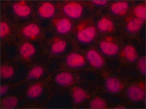
Immunofluorescence Analysis: A representative lot detected RAG-2 in Immunofluorescence applications (Chan, E.A., et. al. (2013). Proc Natl Acad Sci USA. 110(48):E4628-37; Leu, T.M., et. al. (1995). Mol Cell Biol. 15(10):5657-70).

Immunoprecipitation Analysis: A representative lot detected RAG-2 in Immunoprecipitation applications (Leu, T.M., et. al. (1995). Mol Cell Biol. 15(10):5657-70).

Chromatin Immunoprecipitation (ChIP) Analysis: A representative lot detected RAG-2 in Chromatin Immunoprecipitation applications (Dose, M., et. al. (2014). Proc Natl Acad Sci USA. 111(1):391-6; Ji, Y., et. al. (2010). Cell. 141(3):419-31).

Western Blotting Analysis: A representative lot detected RAG-2 in Western Blotting applications (Coster, G., et. al. (2012). J Biol Chem. 287(43):36488-98; Leu, T.M., et. al. (1995). Mol Cell Biol. 15(10):5657-70; Dose, M., et. al. (2014). Proc Natl Acad Sci USA. 111(1):391-6).
Research Category
Epigenetics & Nuclear Function

Quality

Evaluated by Western Blotting in mouse bone marrow tissue lysate.

Western Blotting Analysis: A 1:2,000 dilution of this antibody detected RAG-2 in 10 µg of mouse bone marrow tissue lysate.

Target description

~59 kDa observed; 59.07 kDa calculated. Uncharacterized bands may be observed in some lysate(s).

Physical form

Rabbit monoclonal antibody in tissue culture supernatant without preservatives.
Unpurified

Storage and Stability

Stable for 1 year at -20°C from date of receipt.

Handling Recommendations: Upon receipt and prior to removing the cap, centrifuge the vial and gently mix the solution. Aliquot into microcentrifuge tubes and store at -20°C. Avoid repeated freeze/thaw cycles, which may damage IgG and affect product performance.

Other Notes

Concentration: Please refer to lot specific datasheet.

Disclaimer

Unless otherwise stated in our catalog or other company documentation accompanying the product(s), our products are intended for research use only and are not to be used for any other purpose, which includes but is not limited to, unauthorized commercial uses, in vitro diagnostic uses, ex vivo or in vivo therapeutic uses or any type of consumption or application to humans or animals.

Not finding the right product?  

Try our Product Selector Tool.

Storage Class

10 - Combustible liquids

wgk_germany

WGK 1


Certificates of Analysis (COA)

Search for Certificates of Analysis (COA) by entering the products Lot/Batch Number. Lot and Batch Numbers can be found on a product’s label following the words ‘Lot’ or ‘Batch’.

Already Own This Product?

Find documentation for the products that you have recently purchased in the Document Library.

Visit the Document Library

Our team of scientists has experience in all areas of research including Life Science, Material Science, Chemical Synthesis, Chromatography, Analytical and many others.

Contact Technical Service