Skip to Content
MilliporeSigma
All Photos(1)

Key Documents

MABN483

Sigma-Aldrich

Anti-IRAP Antibody, Clone 3E1

clone 3E1, from mouse

Synonym(s):

Leucyl-cystinyl aminopeptidase, Cystinyl aminopeptidase, Insulin-regulated membrane aminopeptidase, Insulin-responsive aminopeptidase, IRAP, Oxytocinase, OTase, Placental leucine aminopeptidase, P-LAP, Leucyl-cystinyl aminopeptidase, pregnancy serum form

Sign Into View Organizational & Contract Pricing


About This Item

UNSPSC Code:
12352203
eCl@ss:
32160702
NACRES:
NA.41

biological source

mouse

Quality Level

antibody form

purified immunoglobulin

antibody product type

primary antibodies

clone

3E1, monoclonal

species reactivity

human, mouse

technique(s)

immunocytochemistry: suitable
western blot: suitable

isotype

IgG1κ

NCBI accession no.

UniProt accession no.

shipped in

wet ice

target post-translational modification

unmodified

Gene Information

human ... LNPEP(4012)

General description

IRAP, also known as Leucyl-cystinyl aminopeptidase, Cystinyl aminopeptidase, Insulin-regulated membrane aminopeptidase, Insulin-responsive aminopeptidase (IRAP), Oxytocinase (OTase), or Placental leucine aminopeptidase (P-LAP) and encoded by the human gene name IRAP/OTASE, is an important aminopeptidase that plays an important role in maintaining homeostasis during pregnancy. IRAP degrades peptide hormones including oxytocin, vasopressin, and angiotensin III; it also appears to play a role in degrading neuronal peptides in the brain such as Met-enkephalin and dynorphin. IRAP also binds angiotensin IV and appears to be the receptor for angiotensin IV in the brain. IRAP is a membrane bound aminopeptidase and is often found within membrane vesicles that together with other proteins like GLUT4 translocates to the plasma membrane in response to insulin or oxytocin. A cleaved fragment of IRAP, the Leucyl-cystinyl aminopeptidase, is found in the blood during pregnancy, low in the first trimester but rising rapidly until birth, whereby it decreases rapidly, thus the secreted form is often used as a pregnancy marker. IRAP is highly expressed in placenta, heart, kidney and small intestine. IRAP is detected at lower levels in neuronal cells in the brain, in skeletal muscle, spleen, liver, testes and colon.

Immunogen

Epitope: N-terminus
Recombinant protein corresponding to the N-terminus of human IRAP.

Application

Anti-IRAP, Clone 3E1 is an antibody against IRAP for use in WB, ICC.
Research Category
Neuroscience
Research Sub Category
Developmental Signaling
Western Blotting Analysis: A representative lot detected IRAP in 3T3-L1 cells treated with or without insulin (Garza, L.A., et al. (2000). JBC. 275(4):2560-2567).
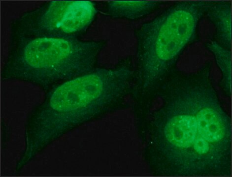
Immunocytochemistry Analysis: A representative lot detected IRAP in 3T3-L1 cells treated with or without insulin (Garza, L.A., et al. (2000). JBC. 275(4):2560-2567).

Quality

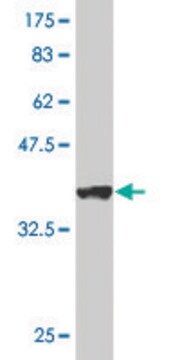
Evaluated by Western Blotting in insulin treated NIH-3T3 cell lysate.

Western Blotting Analysis: 1.0 µg/mL of this antibody detected IRAP in 10 µg of insulin treated NIH-3T3 cell lysate.

Target description

~150 kDa observed. The calculated molecular weight of this protein is 117 kDa, but can be observed at ~150 kDa due to high glycosylation. Uncharacterized band(s) may appear in some lysates.

Physical form

Format: Purified
Protein G Purified
Purified mouse monoclonal IgG1κ in buffer containing 0.1 M Tris-Glycine (pH 7.4), 150 mM NaCl with 0.05% sodium azide.

Storage and Stability

Stable for 1 year at 2-8°C from date of receipt.

Other Notes

Concentration: Please refer to lot specific datasheet.

Disclaimer

Unless otherwise stated in our catalog or other company documentation accompanying the product(s), our products are intended for research use only and are not to be used for any other purpose, which includes but is not limited to, unauthorized commercial uses, in vitro diagnostic uses, ex vivo or in vivo therapeutic uses or any type of consumption or application to humans or animals.

Not finding the right product?  

Try our Product Selector Tool.

Storage Class

12 - Non Combustible Liquids

wgk_germany

WGK 1

flash_point_f

Not applicable

flash_point_c

Not applicable


Certificates of Analysis (COA)

Search for Certificates of Analysis (COA) by entering the products Lot/Batch Number. Lot and Batch Numbers can be found on a product’s label following the words ‘Lot’ or ‘Batch’.

Already Own This Product?

Find documentation for the products that you have recently purchased in the Document Library.

Visit the Document Library

Paula Nunes-Hasler et al.
Nature communications, 8(1), 1852-1852 (2017-11-28)
Antigen cross-presentation by dendritic cells (DC) stimulates cytotoxic T cell activation to promote immunity to intracellular pathogens, viruses and cancer. Phagocytosed antigens generate potent T cell responses, but the signalling and trafficking pathways regulating their cross-presentation are unclear. Here, we

Our team of scientists has experience in all areas of research including Life Science, Material Science, Chemical Synthesis, Chromatography, Analytical and many others.

Contact Technical Service