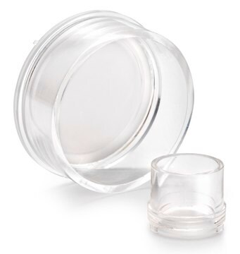
PICM0RG50
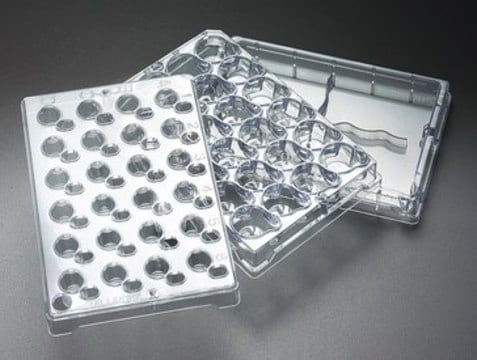
Millicell® Standing Cell Culture Inserts
pore size 0.4 μm, diam. 30 mm, transparent PTFE membrane, hydrophilic, H 5 mm, size 6 wells, sterile
Synonym(s):
Millicell Cell Culture Insert, 30 mm, hydrophilic PTFE, 0.4 µm, Millicell-CM, cell culture inserts, organotypic inserts, plate inserts
About This Item
Recommended Products
material
polystyrene housing
transparent PTFE membrane
Quality Level
sterility
ethylene oxide treated
sterile
feature
hydrophilic
manufacturer/tradename
Millicell®
packaging
pkg of Individually blister packaged
parameter
50 °C max. temp.
technique(s)
cell attachment: suitable
cell culture | mammalian: suitable
cell differentiation: suitable
H
5 mm
diam.
30 mm
size
6 wells
surface area
4.2 cm2
color
transparent membrane, when wetted
matrix
Biopore™
pore size
0.4 μm
binding type
low binding surface
detection method
fluorometric
shipped in
ambient
General description
Application
- Cell Attachment, 3D Cell Culture, Cell Growth, Cell Differentiation , Immunocytochemistry
- Biopore Membrane (hydrophilic PTFE) is ideal for low protein-binding, live cell viewing, and immunofluorescent applications
Features and Benefits
- For high cell viability and superior study of three dimensional explant structure
- Lower height allows them to fit inside a standard petri dish
- The Biopore (PTFE) membrane provides high viability—for as long as 40 days—and excellent trans-membrane oxygen transport
- The membrane is optically clear and optimized for long-term organotypic explant maintenance
Preparation Note
- Add assay solution or media to the receiver well: 2 mL for 6 well format orequivalent dimensions.
- With forceps, place the insert into the receiver well at a 30°-45° angle togradually wet it out from the basolateral side.
- Once edges start to wet out, gently lower the insert until it is fully seatedinto the well.
Legal Information
Storage Class
10-13 - German Storage Class 10 to 13
Certificates of Analysis (COA)
Search for Certificates of Analysis (COA) by entering the products Lot/Batch Number. Lot and Batch Numbers can be found on a product’s label following the words ‘Lot’ or ‘Batch’.
Already Own This Product?
Find documentation for the products that you have recently purchased in the Document Library.
Customers Also Viewed
Protocols
Toluidine blue selectively stains nuclear material and acidic tissue components, aiding in histological staining for tissues rich in DNA/RNA.
This protocol covers 3 modes for the microscopic examination of cell samples.
This page covers the indirect co-culture of embryonic stem cells with embryonic fibroblasts.
This page covers the basic indirect co-culture procedure on both sides of Millicell cell culture insert membranes.
Related Content
Explore our portfolio of 3D cell culture tools and technologies for any 3D cell line or application and discover organoids, hydrogels, ULA, microwell, or hydrogel plates, and more.
Our team of scientists has experience in all areas of research including Life Science, Material Science, Chemical Synthesis, Chromatography, Analytical and many others.
Contact Technical Service