Skip to Content
MilliporeSigma
All Photos(1)

Key Documents

365M-9

Sigma-Aldrich

PNL2 (PNL2) Mouse Monoclonal Antibody

Sign Into View Organizational & Contract Pricing


About This Item

UNSPSC Code:
12352203
NACRES:
NA.41

biological source

mouse

Quality Level

100
500

conjugate

unconjugated

antibody form

culture supernatant

antibody product type

primary antibodies

clone

PNL2, monoclonal

description

For In Vitro Diagnostic Use in Select Regions (See Chart)

form

buffered aqueous solution

species reactivity

human

packaging

vial of 0.1 mL concentrate (365M-94)
vial of 0.5 mL concentrate (365M-95)
bottle of 1.0 mL predilute (365M-97)
vial of 1.0 mL concentrate (365M-96)
bottle of 7.0 mL predilute (365M-98)

manufacturer/tradename

Cell Marque

technique(s)

immunohistochemistry (formalin-fixed, paraffin-embedded sections): 1:25-1:100

isotype

IgG1

control

melanoma

shipped in

wet ice

storage temp.

2-8°C

visualization

cytoplasmic

General description

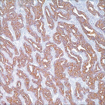
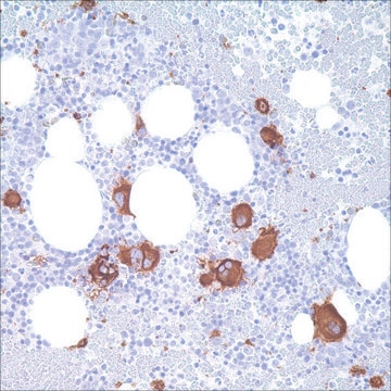
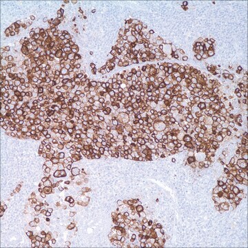
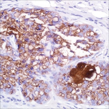
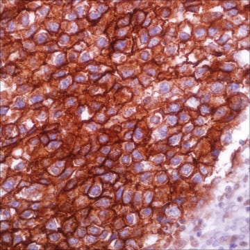
Anti-PNL2 is a novel monoclonal antibody, which has recently been introduced as an immunohistochemical reagent to stain melanocytes and tumors derived therefrom. Anti-PNL2 yields a strong cytoplasmic staining of skin and oral mucosal melanocytes, and staining of granulocytes when used at high concentration. Anti-PNL2 labels intraepidermal nevi while the dermal component of compound nevi are largely non-reactive with anti-PNL2. Antibodies against PNL2, Melan-A and HMB-45 stain most clear cell sarcoma cells and a few cells in angiomyolipomas and lymphangioleiomyomatosis. Non-melanocytic lesions found to be positive with this marker include PEComas and melanotic schwannoma. Anti-PNL2 is a useful antibody for the identification of melanomas and clear cell sarcomas. Differential diagnosis is aided by the results from a panel of antibodies, including antibodies against HMB-45, MART-1, Tyrosinase, and MiTF. The differential diagnosis between benign nevi and malignant melanoma is sometimes difficult, and malignant melanoma may mimic other neoplasms, such as undifferentiated carcinoma, sarcoma or large cell lymphoma. Anti-PNL2 may be most useful because of its high sensitivity for metastatic melanoma (87%), as opposed to 76% for anti-HMB-45 and 82% for anti-MART-1. Furthermore, anti-PNL2, combined with antibodies against S-100 and HMB-45 is a highly sensitive and specific marker for mucosal melanoma.

Quality


IVD

IVD

IVD

RUO

Linkage

PNL2 Positive Control Slides, Product No. 365S, are available for immunohistochemistry (formalin-fixed, paraffin-embedded sections).

Physical form

Solution in Tris Buffer, pH 7.3-7.7, with 1% BSA and <0.1% Sodium Azide

Preparation Note

Download the IFU specific to your product lot and formatNote: This requires a keycode which can be found on your packaging or product label.

Other Notes

For Technical Service please contact: 800-665-7284 or email: service@cellmarque.com

Legal Information

Cell Marque is a trademark of Merck KGaA, Darmstadt, Germany

Not finding the right product?  

Try our Product Selector Tool.


Certificates of Analysis (COA)

Search for Certificates of Analysis (COA) by entering the products Lot/Batch Number. Lot and Batch Numbers can be found on a product’s label following the words ‘Lot’ or ‘Batch’.

Already Own This Product?

Find documentation for the products that you have recently purchased in the Document Library.

Visit the Document Library

Luc G Morris et al.
Head & neck, 30(6), 771-775 (2008-01-30)
Histologic diagnosis of mucosal melanoma of the head and neck is difficult, requiring immunohistochemical stains which are less reliable than in cutaneous lesions. PNL-2 is a novel marker that has not been examined in mucosal melanoma. Nine formalin-fixed tissue sections
Philippe Rochaix et al.
Modern pathology : an official journal of the United States and Canadian Academy of Pathology, Inc, 16(5), 481-490 (2003-05-16)
We report the production of a new monoclonal antibody, PNL2, directed against a fixative resistant melanocyte antigen. The analysis of PNL2 immunostaining on a broad range of normal or malignant human tissues and on various melanocytic lesions revealed its high
Klaus J Busam et al.
The American journal of surgical pathology, 29(3), 400-406 (2005-02-24)
PNL2 is a novel monoclonal antibody, which has recently been introduced as an immunohistochemical reagent to stain melanocyte and tumors derived thereof. In the present study, we analyzed the immunoreactivity of this mAb in various normal tissues, melanocytic nevi, primary
Xiaoning Zhe et al.
The journal of histochemistry and cytochemistry : official journal of the Histochemistry Society, 52(12), 1537-1542 (2004-11-24)
Pulmonary lymphangioleiomyomatosis (LAM) is characterized by abnormal proliferation of immature-looking smooth muscle (SM)-like cells (LAM cells), leading to lung destruction and cyst formation. In addition to expressing some SM markers, scattered LAM cells express the melanocytic maker gp100, which is

Articles

IHC antibodies enhance dermatopathology beyond H&E stained slides, improving techniques and applications for dermatological research.

Our team of scientists has experience in all areas of research including Life Science, Material Science, Chemical Synthesis, Chromatography, Analytical and many others.

Contact Technical Service