Skip to Content
MilliporeSigma
All Photos(3)

Documents

C1478

Sigma-Aldrich

Anti-Calcium Channel (α1B Subunit) (N-type of Voltage-gated Ca2+ Channel) antibody produced in rabbit

affinity isolated antibody, lyophilized powder

Sign Into View Organizational & Contract Pricing


About This Item

MDL number:
UNSPSC Code:
12352203
NACRES:
NA.41

biological source

rabbit

Quality Level

conjugate

unconjugated

antibody form

affinity isolated antibody

antibody product type

primary antibodies

clone

polyclonal

form

lyophilized powder

mol wt

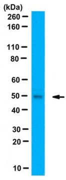
antigen 210 kDa (low)
antigen 240 kDa (high)

species reactivity

rat, mouse

technique(s)

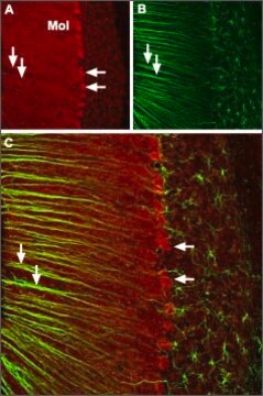
immunocytochemistry: suitable using rat brain sections
immunoprecipitation (IP): suitable
western blot: 1:100-1:200 using rat brain membranes

UniProt accession no.

storage temp.

−20°C

target post-translational modification

unmodified

Gene Information

General description

The calcium channel, voltage-dependent, N type, α1B subunit (CACNA1B) or Cav2.2 gene consists of 47 exons and 46 introns, spanning 244kb. The gene is mapped to human chromosome 9q34. The encoded protein is a member of the voltage-gated calcium channel family.

Immunogen

synthetic peptide corresponding to amino acids 851-867 of the α1B subunit of rat brain voltage-gated calcium channel (VGCC, CNB1), with additional N-terminal lysine and tyrosine, conjugated to KLH.1

Application

Anti-Calcium Channel (α1B Subunit) (N-type of Voltage-gated Ca2+ Channel) antibody produced in rabbit is suitable for the following applications:
  • immunocytochemistry using rat brain sections
  • immunoprecipitation
  • western blotting at a dilution of 1:100-1:200 using rat brain membranes
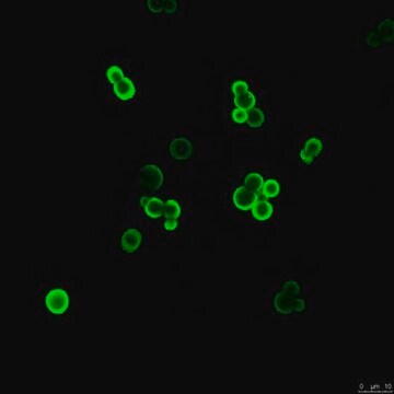
Anti-Calcium Channel (α1B Subunit) (N-type of Voltage-gated Ca2+ Channel) antibody produced in rabbit has been used in immunofluorescence analysis.
Applications in which this antibody has been used successfully, and the associated peer-reviewed papers, are given below.
Immunoprecipitation (1 paper)

Biochem/physiol Actions

Calcium channel, voltage-dependent, N type, α1B subunit (CACNA1B) plays a vital role in regulation of trafficking and biophysical properties of voltage-gated calcium channels. Members of voltage-gated calcium channel family act as key transducers of cell surface membrane potential changes to local intracellular calcium transients that initiate various physiological events. The N-type voltage-gated calcium channels (VGCCs) expressed mainly in the neurons, facilitate neurotransmitter release at the sympathetic nerve terminals. CACNA1B is expressed on presynaptic nerve terminals of nociceptors control neurotransmitter release.

Physical form

Lyophilized from phosphate buffered saline, pH 7.4, containing 1% bovine serum albumin and 0.05% sodium azide.

Disclaimer

Unless otherwise stated in our catalog or other company documentation accompanying the product(s), our products are intended for research use only and are not to be used for any other purpose, which includes but is not limited to, unauthorized commercial uses, in vitro diagnostic uses, ex vivo or in vivo therapeutic uses or any type of consumption or application to humans or animals.

Not finding the right product?  

Try our Product Selector Tool.

Storage Class

11 - Combustible Solids

wgk_germany

WGK 3

flash_point_f

Not applicable

flash_point_c

Not applicable


Certificates of Analysis (COA)

Search for Certificates of Analysis (COA) by entering the products Lot/Batch Number. Lot and Batch Numbers can be found on a product’s label following the words ‘Lot’ or ‘Batch’.

Already Own This Product?

Find documentation for the products that you have recently purchased in the Document Library.

Visit the Document Library

Benjamin Dombert et al.
Frontiers in molecular neuroscience, 10, 346-346 (2017-11-23)
Spontaneous Ca2+ transients and actin dynamics in primary motoneurons correspond to cellular differentiation such as axon elongation and growth cone formation. Brain-derived neurotrophic factor (BDNF) and its receptor trkB support both motoneuron survival and synaptic differentiation. However, in motoneurons effects
Lucian Medrihan et al.
Nature communications, 4, 1512-1512 (2013-02-28)
In the central nervous system, most synapses show a fast mode of neurotransmitter release known as synchronous release followed by a phase of asynchronous release, which extends over tens of milliseconds to seconds. Synapsin II (SYN2) is a member of
Daniele Ferrante et al.
Cell reports, 35(11), 109248-109248 (2021-06-17)
Loss-of-function mutations in proline-rich transmembrane protein-2 (PRRT2) cause paroxysmal disorders associated with defective Ca2+ dependence of glutamatergic transmission. We find that either acute or constitutive PRRT2 deletion induces a significant decrease in the amplitude of evoked excitatory postsynaptic currents (eEPSCs)

Our team of scientists has experience in all areas of research including Life Science, Material Science, Chemical Synthesis, Chromatography, Analytical and many others.

Contact Technical Service